Imrana Tanvir, Sabiha Riaz, Afshan Hussain, Riffat Mehboob, M Usman Shams, Haseeb Ahmad Khan
{"title":"在拉合尔,巴基斯坦的子宫内膜癌上皮恶性肿瘤的频率和常见的诊断缺陷的医院为基础的研究。","authors":"Imrana Tanvir, Sabiha Riaz, Afshan Hussain, Riffat Mehboob, M Usman Shams, Haseeb Ahmad Khan","doi":"10.1155/2014/179384","DOIUrl":null,"url":null,"abstract":"The current study was conducted to see the frequency of epithelial malignancies of endometrium with focus on the common diagnostic pitfalls and identify morphological and immunohistochemical markers helpful in the differential diagnosis between different subtypes. It is a retrospective descriptive study carried out on 52 specimens of endometrial tumors received in Fatima Memorial Hospital, Lahore, Pakistan, during three years (2010–2012). Patients were divided into 5 age groups: <40, 41–50, 51–60, 61–70, and >70 yrs. Tissues were fixed in 10% formalin and processed and stained with haematoxylin-eosin. Stained slides were examined to determine the histological types by WHO classification, and immunohistochemistry for WT1, p53, ER/PR, and MIB1 was done in cases where morphology alone was not helpful in making a confirmed diagnosis. 80% of specimens were of endometrioid adenocarcinomas, 11% of serous tumors, 4% of clear cell carcinoma, and 4% of squamous cell carcinomas involving both cervix and endometrium. Most of the patients (28.84%) with endometrial carcinomas fall in the age range of 51–60 yrs. Endometrioid adenocarcinoma is the most common type of epithelial endometrial malignancies. Morphology is the keystone in the evaluation of these tumors, but immunohistochemistry can also be helpful in establishing the correct diagnosis.","PeriodicalId":89212,"journal":{"name":"Pathology research international","volume":"2014 ","pages":"179384"},"PeriodicalIF":0.0000,"publicationDate":"2014-01-01","publicationTypes":"Journal Article","fieldsOfStudy":null,"isOpenAccess":false,"openAccessPdf":"https://sci-hub-pdf.com/10.1155/2014/179384","citationCount":"12","resultStr":"{\"title\":\"Hospital-based study of epithelial malignancies of endometrial cancer frequency in lahore, pakistan, and common diagnostic pitfalls.\",\"authors\":\"Imrana Tanvir, Sabiha Riaz, Afshan Hussain, Riffat Mehboob, M Usman Shams, Haseeb Ahmad Khan\",\"doi\":\"10.1155/2014/179384\",\"DOIUrl\":null,\"url\":null,\"abstract\":\"The current study was conducted to see the frequency of epithelial malignancies of endometrium with focus on the common diagnostic pitfalls and identify morphological and immunohistochemical markers helpful in the differential diagnosis between different subtypes. It is a retrospective descriptive study carried out on 52 specimens of endometrial tumors received in Fatima Memorial Hospital, Lahore, Pakistan, during three years (2010–2012). Patients were divided into 5 age groups: <40, 41–50, 51–60, 61–70, and >70 yrs. Tissues were fixed in 10% formalin and processed and stained with haematoxylin-eosin. Stained slides were examined to determine the histological types by WHO classification, and immunohistochemistry for WT1, p53, ER/PR, and MIB1 was done in cases where morphology alone was not helpful in making a confirmed diagnosis. 80% of specimens were of endometrioid adenocarcinomas, 11% of serous tumors, 4% of clear cell carcinoma, and 4% of squamous cell carcinomas involving both cervix and endometrium. Most of the patients (28.84%) with endometrial carcinomas fall in the age range of 51–60 yrs. Endometrioid adenocarcinoma is the most common type of epithelial endometrial malignancies. Morphology is the keystone in the evaluation of these tumors, but immunohistochemistry can also be helpful in establishing the correct diagnosis.\",\"PeriodicalId\":89212,\"journal\":{\"name\":\"Pathology research international\",\"volume\":\"2014 \",\"pages\":\"179384\"},\"PeriodicalIF\":0.0000,\"publicationDate\":\"2014-01-01\",\"publicationTypes\":\"Journal Article\",\"fieldsOfStudy\":null,\"isOpenAccess\":false,\"openAccessPdf\":\"https://sci-hub-pdf.com/10.1155/2014/179384\",\"citationCount\":\"12\",\"resultStr\":null,\"platform\":\"Semanticscholar\",\"paperid\":null,\"PeriodicalName\":\"Pathology research international\",\"FirstCategoryId\":\"1085\",\"ListUrlMain\":\"https://doi.org/10.1155/2014/179384\",\"RegionNum\":0,\"RegionCategory\":null,\"ArticlePicture\":[],\"TitleCN\":null,\"AbstractTextCN\":null,\"PMCID\":null,\"EPubDate\":\"2014/1/6 0:00:00\",\"PubModel\":\"Epub\",\"JCR\":\"\",\"JCRName\":\"\",\"Score\":null,\"Total\":0}","platform":"Semanticscholar","paperid":null,"PeriodicalName":"Pathology research international","FirstCategoryId":"1085","ListUrlMain":"https://doi.org/10.1155/2014/179384","RegionNum":0,"RegionCategory":null,"ArticlePicture":[],"TitleCN":null,"AbstractTextCN":null,"PMCID":null,"EPubDate":"2014/1/6 0:00:00","PubModel":"Epub","JCR":"","JCRName":"","Score":null,"Total":0}
Hospital-based study of epithelial malignancies of endometrial cancer frequency in lahore, pakistan, and common diagnostic pitfalls.
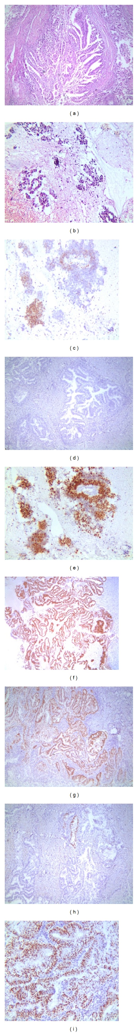
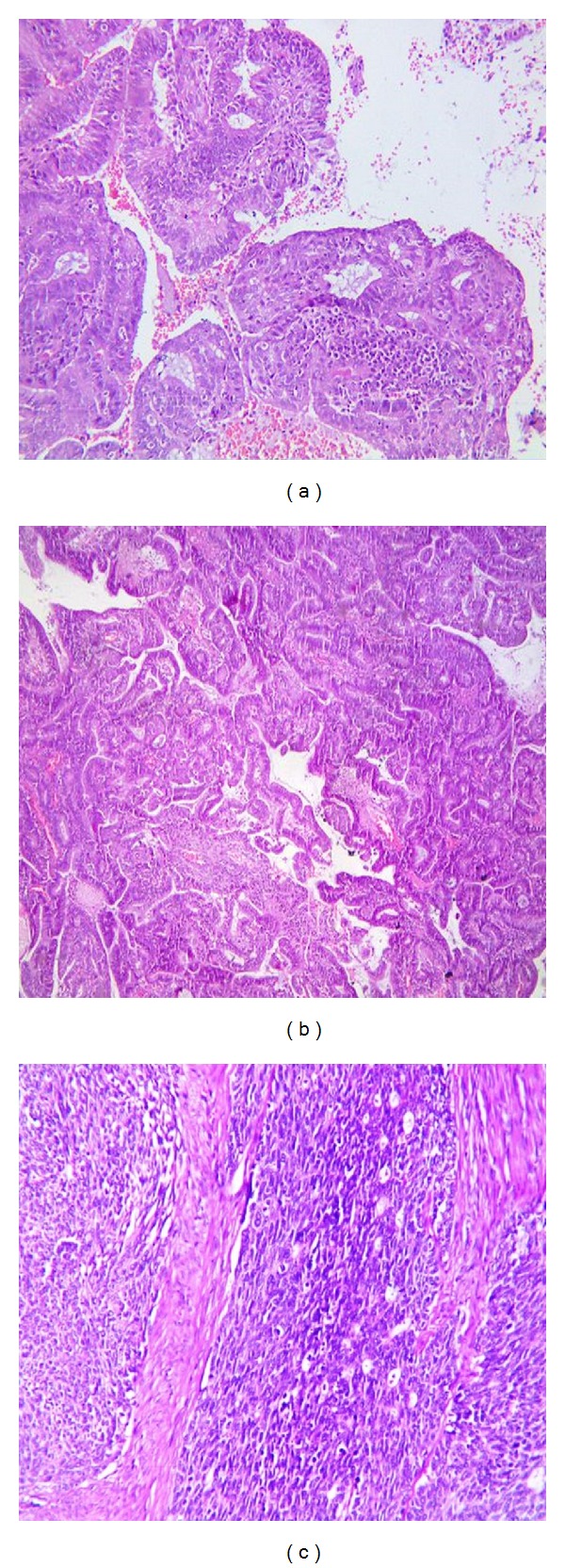
The current study was conducted to see the frequency of epithelial malignancies of endometrium with focus on the common diagnostic pitfalls and identify morphological and immunohistochemical markers helpful in the differential diagnosis between different subtypes. It is a retrospective descriptive study carried out on 52 specimens of endometrial tumors received in Fatima Memorial Hospital, Lahore, Pakistan, during three years (2010–2012). Patients were divided into 5 age groups: <40, 41–50, 51–60, 61–70, and >70 yrs. Tissues were fixed in 10% formalin and processed and stained with haematoxylin-eosin. Stained slides were examined to determine the histological types by WHO classification, and immunohistochemistry for WT1, p53, ER/PR, and MIB1 was done in cases where morphology alone was not helpful in making a confirmed diagnosis. 80% of specimens were of endometrioid adenocarcinomas, 11% of serous tumors, 4% of clear cell carcinoma, and 4% of squamous cell carcinomas involving both cervix and endometrium. Most of the patients (28.84%) with endometrial carcinomas fall in the age range of 51–60 yrs. Endometrioid adenocarcinoma is the most common type of epithelial endometrial malignancies. Morphology is the keystone in the evaluation of these tumors, but immunohistochemistry can also be helpful in establishing the correct diagnosis.

 求助内容:
求助内容: 应助结果提醒方式:


