{"title":"胆管癌体外嗜神经生长的实验研究。","authors":"Yu-Xue Wang, Wei Liu, Xin-Yu Tan, Hui-Huan Tang","doi":"10.1177/2042533313476690","DOIUrl":null,"url":null,"abstract":"<p><strong>Objective: </strong>Perineural invasion of cholangiocarcinoma happens in the early stage of the disease but is often not recognized until its later stages. Research about the behaviour and mechanism of perineural invasion by cholangiocarcinoma is urgently needed for a useful new model. The aim of this work is to establish a novel model to address the problem.</p><p><strong>Design: </strong>Neural cells and cholangiocarcinoma cells were co-cultured to mimic the neurotropic invasion of cholangiocarcinoma.</p><p><strong>Setting: </strong>Human embryonic stem cells were induced to form neural cells by glial cell-derived neurotropic factor and retinoic acid; neural cells and cholangiocarcinoma cells were co-cultured in Transwell chamber.</p><p><strong>Participants: </strong>Human embryonic stem cells and cholangiocarcinoma cells were applied.</p><p><strong>Main outcome measures: </strong>Paired t-test was used to compare the counts of penetrating cholangiocarcinoma cells in co-culture and control group.</p><p><strong>Results: </strong>Formation of neurospheres and neural-like cells were observed following induction at 24 and 48 h, respectively; synapses were viewed to protrude from neural-like cell bodies after incubation for 96 h. Forty-eight hours after incubation, immunocytochemical staining of the cells showed that synaptophysin and glial fibrillary acidic protein were expressed in the neuron-like cells and gliocytes-like cells, respectively. The cholangiocarcinoma cells that had penetrated through the Matrigel/polyethylene terephthalate membrane from the upper chamber to the lower chamber of the Transwell in the co-culture group were significantly more numerous than those in the control group (68 ± 8.3/field versus 46 ± 5.7/field, P < 0.05).</p><p><strong>Conclusion: </strong>The novel model is a valuable tool to study the perineural invasion of cholangiocarcinoma.</p>","PeriodicalId":89182,"journal":{"name":"JRSM short reports","volume":"4 10","pages":"2042533313476690"},"PeriodicalIF":0.0000,"publicationDate":"2013-09-13","publicationTypes":"Journal Article","fieldsOfStudy":null,"isOpenAccess":false,"openAccessPdf":"https://sci-hub-pdf.com/10.1177/2042533313476690","citationCount":"0","resultStr":"{\"title\":\"In vitro neuraotropic growth of cholangiocarcinoma: an experimental study.\",\"authors\":\"Yu-Xue Wang, Wei Liu, Xin-Yu Tan, Hui-Huan Tang\",\"doi\":\"10.1177/2042533313476690\",\"DOIUrl\":null,\"url\":null,\"abstract\":\"<p><strong>Objective: </strong>Perineural invasion of cholangiocarcinoma happens in the early stage of the disease but is often not recognized until its later stages. Research about the behaviour and mechanism of perineural invasion by cholangiocarcinoma is urgently needed for a useful new model. The aim of this work is to establish a novel model to address the problem.</p><p><strong>Design: </strong>Neural cells and cholangiocarcinoma cells were co-cultured to mimic the neurotropic invasion of cholangiocarcinoma.</p><p><strong>Setting: </strong>Human embryonic stem cells were induced to form neural cells by glial cell-derived neurotropic factor and retinoic acid; neural cells and cholangiocarcinoma cells were co-cultured in Transwell chamber.</p><p><strong>Participants: </strong>Human embryonic stem cells and cholangiocarcinoma cells were applied.</p><p><strong>Main outcome measures: </strong>Paired t-test was used to compare the counts of penetrating cholangiocarcinoma cells in co-culture and control group.</p><p><strong>Results: </strong>Formation of neurospheres and neural-like cells were observed following induction at 24 and 48 h, respectively; synapses were viewed to protrude from neural-like cell bodies after incubation for 96 h. Forty-eight hours after incubation, immunocytochemical staining of the cells showed that synaptophysin and glial fibrillary acidic protein were expressed in the neuron-like cells and gliocytes-like cells, respectively. The cholangiocarcinoma cells that had penetrated through the Matrigel/polyethylene terephthalate membrane from the upper chamber to the lower chamber of the Transwell in the co-culture group were significantly more numerous than those in the control group (68 ± 8.3/field versus 46 ± 5.7/field, P < 0.05).</p><p><strong>Conclusion: </strong>The novel model is a valuable tool to study the perineural invasion of cholangiocarcinoma.</p>\",\"PeriodicalId\":89182,\"journal\":{\"name\":\"JRSM short reports\",\"volume\":\"4 10\",\"pages\":\"2042533313476690\"},\"PeriodicalIF\":0.0000,\"publicationDate\":\"2013-09-13\",\"publicationTypes\":\"Journal Article\",\"fieldsOfStudy\":null,\"isOpenAccess\":false,\"openAccessPdf\":\"https://sci-hub-pdf.com/10.1177/2042533313476690\",\"citationCount\":\"0\",\"resultStr\":null,\"platform\":\"Semanticscholar\",\"paperid\":null,\"PeriodicalName\":\"JRSM short reports\",\"FirstCategoryId\":\"1085\",\"ListUrlMain\":\"https://doi.org/10.1177/2042533313476690\",\"RegionNum\":0,\"RegionCategory\":null,\"ArticlePicture\":[],\"TitleCN\":null,\"AbstractTextCN\":null,\"PMCID\":null,\"EPubDate\":\"2013/1/1 0:00:00\",\"PubModel\":\"eCollection\",\"JCR\":\"\",\"JCRName\":\"\",\"Score\":null,\"Total\":0}","platform":"Semanticscholar","paperid":null,"PeriodicalName":"JRSM short reports","FirstCategoryId":"1085","ListUrlMain":"https://doi.org/10.1177/2042533313476690","RegionNum":0,"RegionCategory":null,"ArticlePicture":[],"TitleCN":null,"AbstractTextCN":null,"PMCID":null,"EPubDate":"2013/1/1 0:00:00","PubModel":"eCollection","JCR":"","JCRName":"","Score":null,"Total":0}
In vitro neuraotropic growth of cholangiocarcinoma: an experimental study.
Objective: Perineural invasion of cholangiocarcinoma happens in the early stage of the disease but is often not recognized until its later stages. Research about the behaviour and mechanism of perineural invasion by cholangiocarcinoma is urgently needed for a useful new model. The aim of this work is to establish a novel model to address the problem.
Design: Neural cells and cholangiocarcinoma cells were co-cultured to mimic the neurotropic invasion of cholangiocarcinoma.
Setting: Human embryonic stem cells were induced to form neural cells by glial cell-derived neurotropic factor and retinoic acid; neural cells and cholangiocarcinoma cells were co-cultured in Transwell chamber.
Participants: Human embryonic stem cells and cholangiocarcinoma cells were applied.
Main outcome measures: Paired t-test was used to compare the counts of penetrating cholangiocarcinoma cells in co-culture and control group.
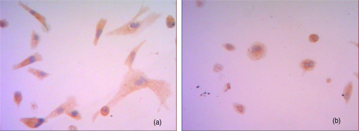
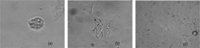
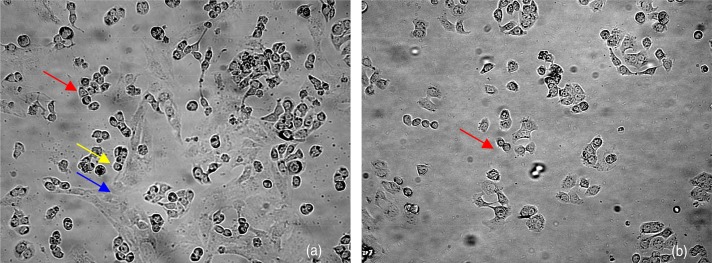
Results: Formation of neurospheres and neural-like cells were observed following induction at 24 and 48 h, respectively; synapses were viewed to protrude from neural-like cell bodies after incubation for 96 h. Forty-eight hours after incubation, immunocytochemical staining of the cells showed that synaptophysin and glial fibrillary acidic protein were expressed in the neuron-like cells and gliocytes-like cells, respectively. The cholangiocarcinoma cells that had penetrated through the Matrigel/polyethylene terephthalate membrane from the upper chamber to the lower chamber of the Transwell in the co-culture group were significantly more numerous than those in the control group (68 ± 8.3/field versus 46 ± 5.7/field, P < 0.05).
Conclusion: The novel model is a valuable tool to study the perineural invasion of cholangiocarcinoma.

 求助内容:
求助内容: 应助结果提醒方式:


