Koon-Chu Yaiw, Olga Ovchinnikova, Chato Taher, Abdul-Aleem Mohammad, Belghis Davoudi, Eugene Shlyakhto, Oxana Rotar, Alexandra Konradi, Vanessa Wilhelmi, Afsar Rahbar, Lynn Butler, Alice Assinger, Cecilia Söderberg-Nauclér
{"title":"俄罗斯颈动脉内膜切除术患者颈动脉粥样硬化斑块中巨细胞病毒的高发率。","authors":"Koon-Chu Yaiw, Olga Ovchinnikova, Chato Taher, Abdul-Aleem Mohammad, Belghis Davoudi, Eugene Shlyakhto, Oxana Rotar, Alexandra Konradi, Vanessa Wilhelmi, Afsar Rahbar, Lynn Butler, Alice Assinger, Cecilia Söderberg-Nauclér","doi":"10.1186/2042-4280-4-3","DOIUrl":null,"url":null,"abstract":"<p><strong>Background: </strong>Human cytomegalovirus (HCMV) infection is associated with cardiovascular disease (CVD) but the role of this virus in CVD progression remains unclear. We aimed to examine the HCMV serostatus in Russian patients (n = 90) who had undergone carotid endarterectomy (CEA) and controls (n = 82) as well as to determine the prevalence of HCMV immediate early (IE) and late (LA) antigens in carotid atherosclerotic plaques obtained from 89 patients. In addition, we sought to determine whether HCMV infection was associated with inflammatory activity in the plaque by quantifying infiltrating CD3 and CD68 positive cells and 5-LO immunoreactivity.</p><p><strong>Methods: </strong>HCMV serology was assessed with ELISA and immunohistochemistry staining was performed to detect HCMV antigens, CD3, CD68 and 5-LO reactivity. The Fisher's exact test was used to compare i) seroprevalence of HCMV IgG between patients and controls and ii) HCMV-positive or -negative to that of CD3, CD68 and 5-LO immunoreactive cells in plaque samples. The student-t test was performed to connote the significance level of mean optical density between patients and controls.</p><p><strong>Results: </strong>The seroprevalence for HCMV IgG was high in both patients and controls (99% and 98%, respectively). Controls had significantly higher IgG titers for HCMV compared with patients (p = 0.0148). Strikingly, we found a high prevalence of HCMV antigens in atherosclerotic plaques; 57/89 (64%) and 47/87 (54%) were HCMV IE and LA positive, respectively. Most plaques had rather low HCMV reactivity with distinct areas of HCMV-positive cells mainly detected in shoulder regions of the plaques, but also in the area adjacent to the necrotic core and fibrous cap. In plaques, the cellular targets for HCMV infection appeared to be mainly macrophages/foam cells and smooth muscle cells. HCMV-positive plaques trended to be associated with increased numbers of CD68 positive macrophages and CD3 positive T cells, while 5-LO reactivity was high in both HCMV-positive and HCMV-negative plaques.</p><p><strong>Conclusions: </strong>In Russian patients undergoing CEA, HCMV proteins are abundantly expressed in carotid plaques and may contribute to the inflammatory response in plaques via enhanced infiltration of CD68 and CD3 cells.</p>","PeriodicalId":89143,"journal":{"name":"Herpesviridae","volume":"4 1","pages":"3"},"PeriodicalIF":0.0000,"publicationDate":"2013-11-14","publicationTypes":"Journal Article","fieldsOfStudy":null,"isOpenAccess":false,"openAccessPdf":"https://sci-hub-pdf.com/10.1186/2042-4280-4-3","citationCount":"17","resultStr":"{\"title\":\"High prevalence of human cytomegalovirus in carotid atherosclerotic plaques obtained from Russian patients undergoing carotid endarterectomy.\",\"authors\":\"Koon-Chu Yaiw, Olga Ovchinnikova, Chato Taher, Abdul-Aleem Mohammad, Belghis Davoudi, Eugene Shlyakhto, Oxana Rotar, Alexandra Konradi, Vanessa Wilhelmi, Afsar Rahbar, Lynn Butler, Alice Assinger, Cecilia Söderberg-Nauclér\",\"doi\":\"10.1186/2042-4280-4-3\",\"DOIUrl\":null,\"url\":null,\"abstract\":\"<p><strong>Background: </strong>Human cytomegalovirus (HCMV) infection is associated with cardiovascular disease (CVD) but the role of this virus in CVD progression remains unclear. We aimed to examine the HCMV serostatus in Russian patients (n = 90) who had undergone carotid endarterectomy (CEA) and controls (n = 82) as well as to determine the prevalence of HCMV immediate early (IE) and late (LA) antigens in carotid atherosclerotic plaques obtained from 89 patients. In addition, we sought to determine whether HCMV infection was associated with inflammatory activity in the plaque by quantifying infiltrating CD3 and CD68 positive cells and 5-LO immunoreactivity.</p><p><strong>Methods: </strong>HCMV serology was assessed with ELISA and immunohistochemistry staining was performed to detect HCMV antigens, CD3, CD68 and 5-LO reactivity. The Fisher's exact test was used to compare i) seroprevalence of HCMV IgG between patients and controls and ii) HCMV-positive or -negative to that of CD3, CD68 and 5-LO immunoreactive cells in plaque samples. The student-t test was performed to connote the significance level of mean optical density between patients and controls.</p><p><strong>Results: </strong>The seroprevalence for HCMV IgG was high in both patients and controls (99% and 98%, respectively). Controls had significantly higher IgG titers for HCMV compared with patients (p = 0.0148). Strikingly, we found a high prevalence of HCMV antigens in atherosclerotic plaques; 57/89 (64%) and 47/87 (54%) were HCMV IE and LA positive, respectively. Most plaques had rather low HCMV reactivity with distinct areas of HCMV-positive cells mainly detected in shoulder regions of the plaques, but also in the area adjacent to the necrotic core and fibrous cap. In plaques, the cellular targets for HCMV infection appeared to be mainly macrophages/foam cells and smooth muscle cells. HCMV-positive plaques trended to be associated with increased numbers of CD68 positive macrophages and CD3 positive T cells, while 5-LO reactivity was high in both HCMV-positive and HCMV-negative plaques.</p><p><strong>Conclusions: </strong>In Russian patients undergoing CEA, HCMV proteins are abundantly expressed in carotid plaques and may contribute to the inflammatory response in plaques via enhanced infiltration of CD68 and CD3 cells.</p>\",\"PeriodicalId\":89143,\"journal\":{\"name\":\"Herpesviridae\",\"volume\":\"4 1\",\"pages\":\"3\"},\"PeriodicalIF\":0.0000,\"publicationDate\":\"2013-11-14\",\"publicationTypes\":\"Journal Article\",\"fieldsOfStudy\":null,\"isOpenAccess\":false,\"openAccessPdf\":\"https://sci-hub-pdf.com/10.1186/2042-4280-4-3\",\"citationCount\":\"17\",\"resultStr\":null,\"platform\":\"Semanticscholar\",\"paperid\":null,\"PeriodicalName\":\"Herpesviridae\",\"FirstCategoryId\":\"1085\",\"ListUrlMain\":\"https://doi.org/10.1186/2042-4280-4-3\",\"RegionNum\":0,\"RegionCategory\":null,\"ArticlePicture\":[],\"TitleCN\":null,\"AbstractTextCN\":null,\"PMCID\":null,\"EPubDate\":\"\",\"PubModel\":\"\",\"JCR\":\"\",\"JCRName\":\"\",\"Score\":null,\"Total\":0}","platform":"Semanticscholar","paperid":null,"PeriodicalName":"Herpesviridae","FirstCategoryId":"1085","ListUrlMain":"https://doi.org/10.1186/2042-4280-4-3","RegionNum":0,"RegionCategory":null,"ArticlePicture":[],"TitleCN":null,"AbstractTextCN":null,"PMCID":null,"EPubDate":"","PubModel":"","JCR":"","JCRName":"","Score":null,"Total":0}
High prevalence of human cytomegalovirus in carotid atherosclerotic plaques obtained from Russian patients undergoing carotid endarterectomy.
Background: Human cytomegalovirus (HCMV) infection is associated with cardiovascular disease (CVD) but the role of this virus in CVD progression remains unclear. We aimed to examine the HCMV serostatus in Russian patients (n = 90) who had undergone carotid endarterectomy (CEA) and controls (n = 82) as well as to determine the prevalence of HCMV immediate early (IE) and late (LA) antigens in carotid atherosclerotic plaques obtained from 89 patients. In addition, we sought to determine whether HCMV infection was associated with inflammatory activity in the plaque by quantifying infiltrating CD3 and CD68 positive cells and 5-LO immunoreactivity.
Methods: HCMV serology was assessed with ELISA and immunohistochemistry staining was performed to detect HCMV antigens, CD3, CD68 and 5-LO reactivity. The Fisher's exact test was used to compare i) seroprevalence of HCMV IgG between patients and controls and ii) HCMV-positive or -negative to that of CD3, CD68 and 5-LO immunoreactive cells in plaque samples. The student-t test was performed to connote the significance level of mean optical density between patients and controls.
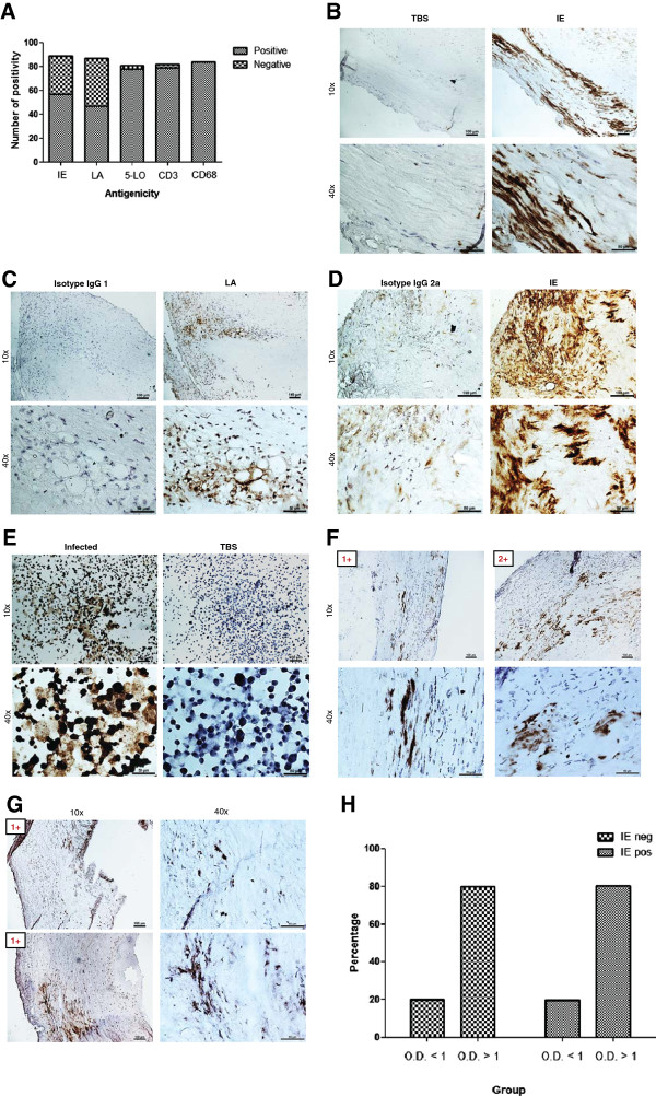
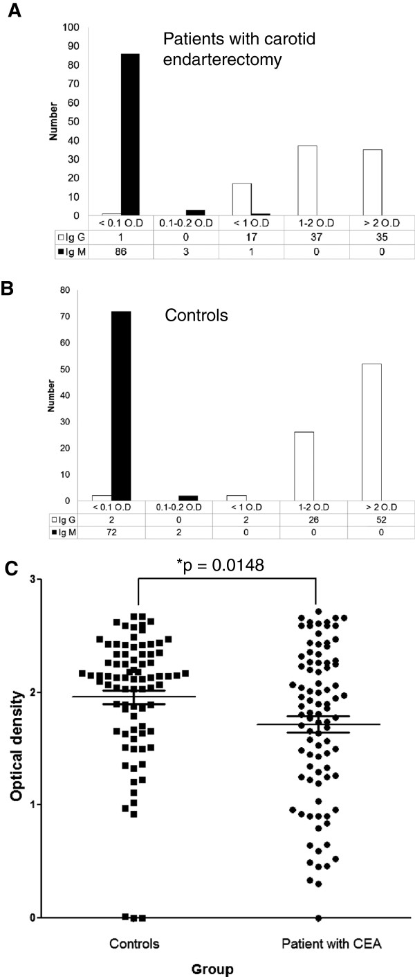
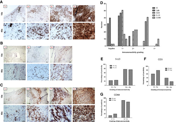
Results: The seroprevalence for HCMV IgG was high in both patients and controls (99% and 98%, respectively). Controls had significantly higher IgG titers for HCMV compared with patients (p = 0.0148). Strikingly, we found a high prevalence of HCMV antigens in atherosclerotic plaques; 57/89 (64%) and 47/87 (54%) were HCMV IE and LA positive, respectively. Most plaques had rather low HCMV reactivity with distinct areas of HCMV-positive cells mainly detected in shoulder regions of the plaques, but also in the area adjacent to the necrotic core and fibrous cap. In plaques, the cellular targets for HCMV infection appeared to be mainly macrophages/foam cells and smooth muscle cells. HCMV-positive plaques trended to be associated with increased numbers of CD68 positive macrophages and CD3 positive T cells, while 5-LO reactivity was high in both HCMV-positive and HCMV-negative plaques.
Conclusions: In Russian patients undergoing CEA, HCMV proteins are abundantly expressed in carotid plaques and may contribute to the inflammatory response in plaques via enhanced infiltration of CD68 and CD3 cells.

 求助内容:
求助内容: 应助结果提醒方式:


