Geeta Devi Leishangthem, Ulaganathan Mabalirajan, Vijay Pal Singh, Anurag Agrawal, Balaram Ghosh, Amit Kumar Dinda
{"title":"过敏和饮食诱发代谢综合征小鼠模型的气道超微结构变化。","authors":"Geeta Devi Leishangthem, Ulaganathan Mabalirajan, Vijay Pal Singh, Anurag Agrawal, Balaram Ghosh, Amit Kumar Dinda","doi":"10.1155/2013/261297","DOIUrl":null,"url":null,"abstract":"<p><p>Studying ultrastructural changes could reveal novel pathophysiology of obese-asthmatic condition as existing concepts in asthma pathogenesis are based on the histological changes of the diseased airway. While asthma is defined in functional terms, the potential of electron microscopy (EM) in providing cellular and subcellular detail is underutilized. With this view, we have performed transmission EM in the lungs from allergic mice that show key features of asthma and high-fat- or high-fructose-fed mice that mimicked metabolic syndrome to illustrate the ultrastructural changes. The primary focus was epithelial injury and metaplasia, which are cardinal features of asthma and initiate airway remodeling. EM findings of the allergically inflamed mouse lungs correlate with known features of human asthma such as increased mitochondria in airway smooth muscle, platelet activation and subepithelial myofibroblasts. Interestingly, we found a clear and unambiguous evidence to suggest that ciliated cells can become goblet cells using immunoelectron microscopy. Additionally, we show for the first time the stressed mitochondria in the bronchial epithelia of high-fat- or high-fructose-fed mice even without allergen exposure. These results may stimulate interest in using EM in understanding novel pathological mechanisms for different subtypes of asthma including obese asthma. </p>","PeriodicalId":89782,"journal":{"name":"ISRN allergy","volume":"2013 ","pages":"261297"},"PeriodicalIF":0.0000,"publicationDate":"2013-09-10","publicationTypes":"Journal Article","fieldsOfStudy":null,"isOpenAccess":false,"openAccessPdf":"https://www.ncbi.nlm.nih.gov/pmc/articles/PMC3782840/pdf/","citationCount":"0","resultStr":"{\"title\":\"Ultrastructural changes of airway in murine models of allergy and diet-induced metabolic syndrome.\",\"authors\":\"Geeta Devi Leishangthem, Ulaganathan Mabalirajan, Vijay Pal Singh, Anurag Agrawal, Balaram Ghosh, Amit Kumar Dinda\",\"doi\":\"10.1155/2013/261297\",\"DOIUrl\":null,\"url\":null,\"abstract\":\"<p><p>Studying ultrastructural changes could reveal novel pathophysiology of obese-asthmatic condition as existing concepts in asthma pathogenesis are based on the histological changes of the diseased airway. While asthma is defined in functional terms, the potential of electron microscopy (EM) in providing cellular and subcellular detail is underutilized. With this view, we have performed transmission EM in the lungs from allergic mice that show key features of asthma and high-fat- or high-fructose-fed mice that mimicked metabolic syndrome to illustrate the ultrastructural changes. The primary focus was epithelial injury and metaplasia, which are cardinal features of asthma and initiate airway remodeling. EM findings of the allergically inflamed mouse lungs correlate with known features of human asthma such as increased mitochondria in airway smooth muscle, platelet activation and subepithelial myofibroblasts. Interestingly, we found a clear and unambiguous evidence to suggest that ciliated cells can become goblet cells using immunoelectron microscopy. Additionally, we show for the first time the stressed mitochondria in the bronchial epithelia of high-fat- or high-fructose-fed mice even without allergen exposure. These results may stimulate interest in using EM in understanding novel pathological mechanisms for different subtypes of asthma including obese asthma. </p>\",\"PeriodicalId\":89782,\"journal\":{\"name\":\"ISRN allergy\",\"volume\":\"2013 \",\"pages\":\"261297\"},\"PeriodicalIF\":0.0000,\"publicationDate\":\"2013-09-10\",\"publicationTypes\":\"Journal Article\",\"fieldsOfStudy\":null,\"isOpenAccess\":false,\"openAccessPdf\":\"https://www.ncbi.nlm.nih.gov/pmc/articles/PMC3782840/pdf/\",\"citationCount\":\"0\",\"resultStr\":null,\"platform\":\"Semanticscholar\",\"paperid\":null,\"PeriodicalName\":\"ISRN allergy\",\"FirstCategoryId\":\"1085\",\"ListUrlMain\":\"https://doi.org/10.1155/2013/261297\",\"RegionNum\":0,\"RegionCategory\":null,\"ArticlePicture\":[],\"TitleCN\":null,\"AbstractTextCN\":null,\"PMCID\":null,\"EPubDate\":\"2013/1/1 0:00:00\",\"PubModel\":\"eCollection\",\"JCR\":\"\",\"JCRName\":\"\",\"Score\":null,\"Total\":0}","platform":"Semanticscholar","paperid":null,"PeriodicalName":"ISRN allergy","FirstCategoryId":"1085","ListUrlMain":"https://doi.org/10.1155/2013/261297","RegionNum":0,"RegionCategory":null,"ArticlePicture":[],"TitleCN":null,"AbstractTextCN":null,"PMCID":null,"EPubDate":"2013/1/1 0:00:00","PubModel":"eCollection","JCR":"","JCRName":"","Score":null,"Total":0}
Ultrastructural changes of airway in murine models of allergy and diet-induced metabolic syndrome.
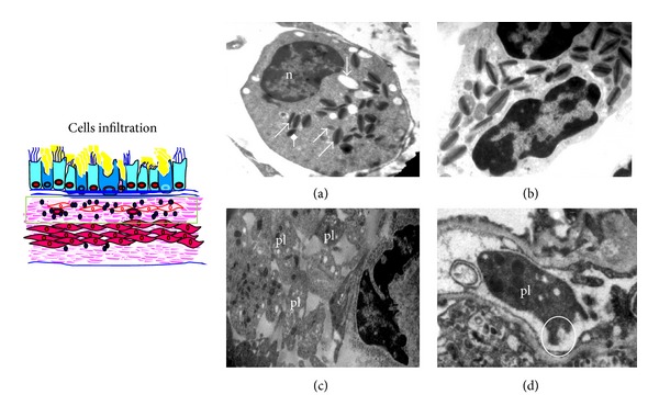
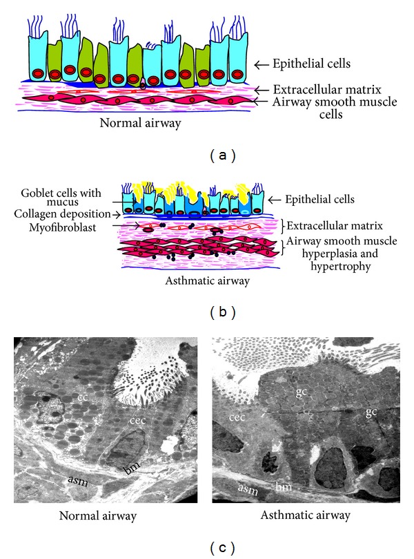
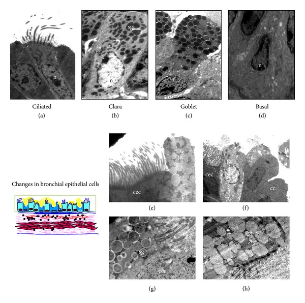
Studying ultrastructural changes could reveal novel pathophysiology of obese-asthmatic condition as existing concepts in asthma pathogenesis are based on the histological changes of the diseased airway. While asthma is defined in functional terms, the potential of electron microscopy (EM) in providing cellular and subcellular detail is underutilized. With this view, we have performed transmission EM in the lungs from allergic mice that show key features of asthma and high-fat- or high-fructose-fed mice that mimicked metabolic syndrome to illustrate the ultrastructural changes. The primary focus was epithelial injury and metaplasia, which are cardinal features of asthma and initiate airway remodeling. EM findings of the allergically inflamed mouse lungs correlate with known features of human asthma such as increased mitochondria in airway smooth muscle, platelet activation and subepithelial myofibroblasts. Interestingly, we found a clear and unambiguous evidence to suggest that ciliated cells can become goblet cells using immunoelectron microscopy. Additionally, we show for the first time the stressed mitochondria in the bronchial epithelia of high-fat- or high-fructose-fed mice even without allergen exposure. These results may stimulate interest in using EM in understanding novel pathological mechanisms for different subtypes of asthma including obese asthma.

 求助内容:
求助内容: 应助结果提醒方式:


