{"title":"在资源有限的国家,超声检查和乳房x光检查对乳房肿块的癌症调查是必要的吗?","authors":"Rungnapa Chairat, Adisorn Puttisri, Asani Pamarapa, Sahatham Samintharapanya, Chamaiporn Tawichasri, Jayanton Patumanond","doi":"10.1155/2013/257942","DOIUrl":null,"url":null,"abstract":"<p><p>Objective. To reevaluate the diagnostic value of breast imaging in the diagnosis of breast cancer in areas where health resources are limited. Methods. Patients were women presenting with breast lumps in two university-affiliated tertiary hospitals, Thailand, during 2006 and 2010. Clinical data were abstracted from the breast cancer registration database and patient records. The diagnostic predictive ability of ultrasonography and mammography was obtained from logistic regression analysis and presented with areas under the receiver operating characteristics (AuROCs) curves. Results. Among 3129 breast lumps (3069 women), 854 were diagnosed with breast cancer by certified pathologists. Age and size of lumps alone already predicted cancer correctly in 77.45% (AuROC = 77.45). Additional ultrasonography increased the prediction to 96.22% (P < 0.001). Additional mammography also increased the prediction to 95.99% (P < 0.001). Performing both imaging modalities did not increase the prediction clinically (0.01%-0.24%). More accurate prediction (2.07%-2.21%) may be added by fine needle aspiration cytology (FNAC). Conclusions. Breast imaging is still valuable in settings where health resources are limited. Single breast imaging (only either ultrasonography or mammography) is adequate for cancer diagnosis. It is therefore unnecessary to perform both imaging modalities. Accuracy of the diagnosis may be improved by FNAC, if available. </p>","PeriodicalId":89399,"journal":{"name":"ISRN oncology","volume":"2013 ","pages":"257942"},"PeriodicalIF":0.0000,"publicationDate":"2013-08-28","publicationTypes":"Journal Article","fieldsOfStudy":null,"isOpenAccess":false,"openAccessPdf":"https://sci-hub-pdf.com/10.1155/2013/257942","citationCount":"9","resultStr":"{\"title\":\"Are both ultrasonography and mammography necessary for cancer investigation of breast lumps in resource-limited countries?\",\"authors\":\"Rungnapa Chairat, Adisorn Puttisri, Asani Pamarapa, Sahatham Samintharapanya, Chamaiporn Tawichasri, Jayanton Patumanond\",\"doi\":\"10.1155/2013/257942\",\"DOIUrl\":null,\"url\":null,\"abstract\":\"<p><p>Objective. To reevaluate the diagnostic value of breast imaging in the diagnosis of breast cancer in areas where health resources are limited. Methods. Patients were women presenting with breast lumps in two university-affiliated tertiary hospitals, Thailand, during 2006 and 2010. Clinical data were abstracted from the breast cancer registration database and patient records. The diagnostic predictive ability of ultrasonography and mammography was obtained from logistic regression analysis and presented with areas under the receiver operating characteristics (AuROCs) curves. Results. Among 3129 breast lumps (3069 women), 854 were diagnosed with breast cancer by certified pathologists. Age and size of lumps alone already predicted cancer correctly in 77.45% (AuROC = 77.45). Additional ultrasonography increased the prediction to 96.22% (P < 0.001). Additional mammography also increased the prediction to 95.99% (P < 0.001). Performing both imaging modalities did not increase the prediction clinically (0.01%-0.24%). More accurate prediction (2.07%-2.21%) may be added by fine needle aspiration cytology (FNAC). Conclusions. Breast imaging is still valuable in settings where health resources are limited. Single breast imaging (only either ultrasonography or mammography) is adequate for cancer diagnosis. It is therefore unnecessary to perform both imaging modalities. Accuracy of the diagnosis may be improved by FNAC, if available. </p>\",\"PeriodicalId\":89399,\"journal\":{\"name\":\"ISRN oncology\",\"volume\":\"2013 \",\"pages\":\"257942\"},\"PeriodicalIF\":0.0000,\"publicationDate\":\"2013-08-28\",\"publicationTypes\":\"Journal Article\",\"fieldsOfStudy\":null,\"isOpenAccess\":false,\"openAccessPdf\":\"https://sci-hub-pdf.com/10.1155/2013/257942\",\"citationCount\":\"9\",\"resultStr\":null,\"platform\":\"Semanticscholar\",\"paperid\":null,\"PeriodicalName\":\"ISRN oncology\",\"FirstCategoryId\":\"1085\",\"ListUrlMain\":\"https://doi.org/10.1155/2013/257942\",\"RegionNum\":0,\"RegionCategory\":null,\"ArticlePicture\":[],\"TitleCN\":null,\"AbstractTextCN\":null,\"PMCID\":null,\"EPubDate\":\"2013/1/1 0:00:00\",\"PubModel\":\"eCollection\",\"JCR\":\"\",\"JCRName\":\"\",\"Score\":null,\"Total\":0}","platform":"Semanticscholar","paperid":null,"PeriodicalName":"ISRN oncology","FirstCategoryId":"1085","ListUrlMain":"https://doi.org/10.1155/2013/257942","RegionNum":0,"RegionCategory":null,"ArticlePicture":[],"TitleCN":null,"AbstractTextCN":null,"PMCID":null,"EPubDate":"2013/1/1 0:00:00","PubModel":"eCollection","JCR":"","JCRName":"","Score":null,"Total":0}
Are both ultrasonography and mammography necessary for cancer investigation of breast lumps in resource-limited countries?
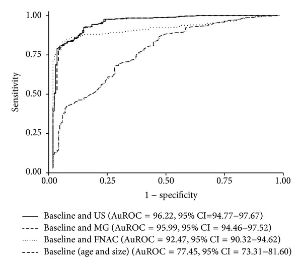
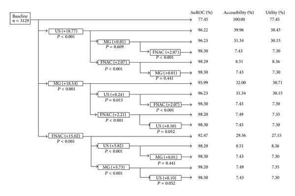
Objective. To reevaluate the diagnostic value of breast imaging in the diagnosis of breast cancer in areas where health resources are limited. Methods. Patients were women presenting with breast lumps in two university-affiliated tertiary hospitals, Thailand, during 2006 and 2010. Clinical data were abstracted from the breast cancer registration database and patient records. The diagnostic predictive ability of ultrasonography and mammography was obtained from logistic regression analysis and presented with areas under the receiver operating characteristics (AuROCs) curves. Results. Among 3129 breast lumps (3069 women), 854 were diagnosed with breast cancer by certified pathologists. Age and size of lumps alone already predicted cancer correctly in 77.45% (AuROC = 77.45). Additional ultrasonography increased the prediction to 96.22% (P < 0.001). Additional mammography also increased the prediction to 95.99% (P < 0.001). Performing both imaging modalities did not increase the prediction clinically (0.01%-0.24%). More accurate prediction (2.07%-2.21%) may be added by fine needle aspiration cytology (FNAC). Conclusions. Breast imaging is still valuable in settings where health resources are limited. Single breast imaging (only either ultrasonography or mammography) is adequate for cancer diagnosis. It is therefore unnecessary to perform both imaging modalities. Accuracy of the diagnosis may be improved by FNAC, if available.

 求助内容:
求助内容: 应助结果提醒方式:


