Fabio Tavora, Mingchang Zhang, Nathaniel Cresswell, Ling Li, David Fowler, Marcello Franco, Allen Burke
{"title":"致心律失常心肌病中桥粒体蛋白(血小板红蛋白、桥粒蛋白和嗜血小板蛋白)、连接蛋白-43和n-钙粘蛋白的定量免疫组化:一项尸检研究。","authors":"Fabio Tavora, Mingchang Zhang, Nathaniel Cresswell, Ling Li, David Fowler, Marcello Franco, Allen Burke","doi":"10.2174/1874192401307010028","DOIUrl":null,"url":null,"abstract":"<p><strong>Background: </strong>Arrhythmogenic right ventricular cardiomyopathy (ARVC) is a genetic disorder related to mutations in desmosomal proteins. The current study tests the hypothesis that immunohistochemical staining for desmosomal proteins is of diagnostic utility by studying autopsy-confirmed cases of ARVC.</p><p><strong>Methods and results: </strong>We studied 23 hearts from patients dying suddenly with ARVC. Control subject tissues were 21 hearts from people dying from non-cardiac causes (n=15), dilated cardiomyopathy (n=3) and coronary artery disease (n=3). Areas free of fibrofatty change or scarring were assessed on 50 sections from ARVC (24 left ventricle, 26 right ventricle) and 28 sections from controls. Immunohistochemical stains against plakoglobin, plakophilin, desmoplakin, connexin-43, and N-cadherin were applied and area expression analyzed by computerized morphometry. Desmin was stained as a control for fixation and similarly analyzed. The mean area of desmin expression was similar in controls and ARVC (86% vs. 85%, p=0.6). Plakoglobin expression was 4.9% ± 0.3% in controls, vs. 4.6% ± 0.3% in ARVC (p=0.3). Plakophilin staining was 4.8% ± 0.3% in controls vs. 4.4% ± 03% in ARVC (p=0.3). Desmoplakin staining was 3.4% in controls vs. 3.2 ± 0.2% in ARVC (p=0.6). There were no significant differences when staining was compared between right and left ventricles (all p > 0.1). For non-desmosomal proteins, the mean area of connexin-43 staining showed no significant difference by presence of disease.</p><p><strong>Conclusions: </strong>The small and insignificant decrease in junction protein expression in ARVC suggests that immunohistochemistry is not a useful tool for the diagnosis.</p>","PeriodicalId":504447,"journal":{"name":"The Open Cardiovascular Medicine Journal","volume":"7 ","pages":"28-35"},"PeriodicalIF":0.0000,"publicationDate":"2013-03-29","publicationTypes":"Journal Article","fieldsOfStudy":null,"isOpenAccess":false,"openAccessPdf":"https://ftp.ncbi.nlm.nih.gov/pub/pmc/oa_pdf/fb/e2/TOCMJ-7-28.PMC3680985.pdf","citationCount":"11","resultStr":"{\"title\":\"Quantitative Immunohistochemistry of Desmosomal Proteins (Plakoglobin, Desmoplakin and Plakophilin), Connexin-43, and N-cadherin in Arrhythmogenic Cardiomyopathy: An Autopsy Study.\",\"authors\":\"Fabio Tavora, Mingchang Zhang, Nathaniel Cresswell, Ling Li, David Fowler, Marcello Franco, Allen Burke\",\"doi\":\"10.2174/1874192401307010028\",\"DOIUrl\":null,\"url\":null,\"abstract\":\"<p><strong>Background: </strong>Arrhythmogenic right ventricular cardiomyopathy (ARVC) is a genetic disorder related to mutations in desmosomal proteins. The current study tests the hypothesis that immunohistochemical staining for desmosomal proteins is of diagnostic utility by studying autopsy-confirmed cases of ARVC.</p><p><strong>Methods and results: </strong>We studied 23 hearts from patients dying suddenly with ARVC. Control subject tissues were 21 hearts from people dying from non-cardiac causes (n=15), dilated cardiomyopathy (n=3) and coronary artery disease (n=3). Areas free of fibrofatty change or scarring were assessed on 50 sections from ARVC (24 left ventricle, 26 right ventricle) and 28 sections from controls. Immunohistochemical stains against plakoglobin, plakophilin, desmoplakin, connexin-43, and N-cadherin were applied and area expression analyzed by computerized morphometry. Desmin was stained as a control for fixation and similarly analyzed. The mean area of desmin expression was similar in controls and ARVC (86% vs. 85%, p=0.6). Plakoglobin expression was 4.9% ± 0.3% in controls, vs. 4.6% ± 0.3% in ARVC (p=0.3). Plakophilin staining was 4.8% ± 0.3% in controls vs. 4.4% ± 03% in ARVC (p=0.3). Desmoplakin staining was 3.4% in controls vs. 3.2 ± 0.2% in ARVC (p=0.6). There were no significant differences when staining was compared between right and left ventricles (all p > 0.1). For non-desmosomal proteins, the mean area of connexin-43 staining showed no significant difference by presence of disease.</p><p><strong>Conclusions: </strong>The small and insignificant decrease in junction protein expression in ARVC suggests that immunohistochemistry is not a useful tool for the diagnosis.</p>\",\"PeriodicalId\":504447,\"journal\":{\"name\":\"The Open Cardiovascular Medicine Journal\",\"volume\":\"7 \",\"pages\":\"28-35\"},\"PeriodicalIF\":0.0000,\"publicationDate\":\"2013-03-29\",\"publicationTypes\":\"Journal Article\",\"fieldsOfStudy\":null,\"isOpenAccess\":false,\"openAccessPdf\":\"https://ftp.ncbi.nlm.nih.gov/pub/pmc/oa_pdf/fb/e2/TOCMJ-7-28.PMC3680985.pdf\",\"citationCount\":\"11\",\"resultStr\":null,\"platform\":\"Semanticscholar\",\"paperid\":null,\"PeriodicalName\":\"The Open Cardiovascular Medicine Journal\",\"FirstCategoryId\":\"1085\",\"ListUrlMain\":\"https://doi.org/10.2174/1874192401307010028\",\"RegionNum\":0,\"RegionCategory\":null,\"ArticlePicture\":[],\"TitleCN\":null,\"AbstractTextCN\":null,\"PMCID\":null,\"EPubDate\":\"2013/1/1 0:00:00\",\"PubModel\":\"Print\",\"JCR\":\"\",\"JCRName\":\"\",\"Score\":null,\"Total\":0}","platform":"Semanticscholar","paperid":null,"PeriodicalName":"The Open Cardiovascular Medicine Journal","FirstCategoryId":"1085","ListUrlMain":"https://doi.org/10.2174/1874192401307010028","RegionNum":0,"RegionCategory":null,"ArticlePicture":[],"TitleCN":null,"AbstractTextCN":null,"PMCID":null,"EPubDate":"2013/1/1 0:00:00","PubModel":"Print","JCR":"","JCRName":"","Score":null,"Total":0}
引用次数: 11
摘要
背景:心律失常性右室心肌病(ARVC)是一种与桥粒体蛋白突变有关的遗传性疾病。目前的研究通过研究尸检确诊的ARVC病例,验证了桥粒蛋白免疫组织化学染色具有诊断作用的假设。方法与结果:对23例猝死ARVC患者的心脏进行研究。对照受试者组织为21颗心脏,分别来自非心脏原因死亡患者(n=15)、扩张型心肌病患者(n=3)和冠状动脉疾病患者(n=3)。在ARVC的50个切片(24个左心室,26个右心室)和28个对照组切片上评估无纤维脂肪改变或瘢痕形成的区域。应用血小板红蛋白、嗜血小板蛋白、桥蛋白、连接蛋白-43和n-钙粘蛋白免疫组化染色,并用计算机形态测定法分析面积表达。Desmin染色作为固定对照,并进行类似分析。在对照组和ARVC中desmin的平均表达面积相似(86% vs. 85%, p=0.6)。对照组血小板红蛋白表达为4.9%±0.3%,ARVC组为4.6%±0.3% (p=0.3)。嗜白细胞染色在对照组为4.8%±0.3%,在ARVC组为4.4%±03% (p=0.3)。在对照组中,桥蛋白染色为3.4%,在ARVC中为3.2±0.2% (p=0.6)。左、右心室染色比较差异无统计学意义(p > 0.1)。对于非桥粒体蛋白,连接蛋白-43染色的平均面积没有显示出疾病存在的显著差异。结论:ARVC中连接蛋白表达的微小且不显著的下降提示免疫组化不是诊断ARVC的有效工具。
Quantitative Immunohistochemistry of Desmosomal Proteins (Plakoglobin, Desmoplakin and Plakophilin), Connexin-43, and N-cadherin in Arrhythmogenic Cardiomyopathy: An Autopsy Study.
Background: Arrhythmogenic right ventricular cardiomyopathy (ARVC) is a genetic disorder related to mutations in desmosomal proteins. The current study tests the hypothesis that immunohistochemical staining for desmosomal proteins is of diagnostic utility by studying autopsy-confirmed cases of ARVC.
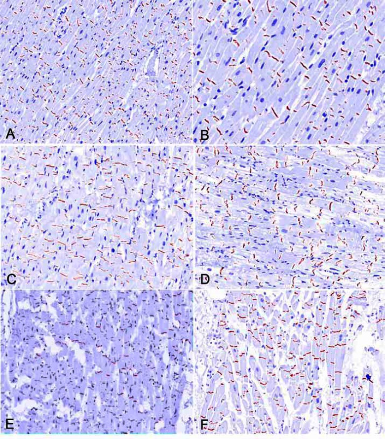
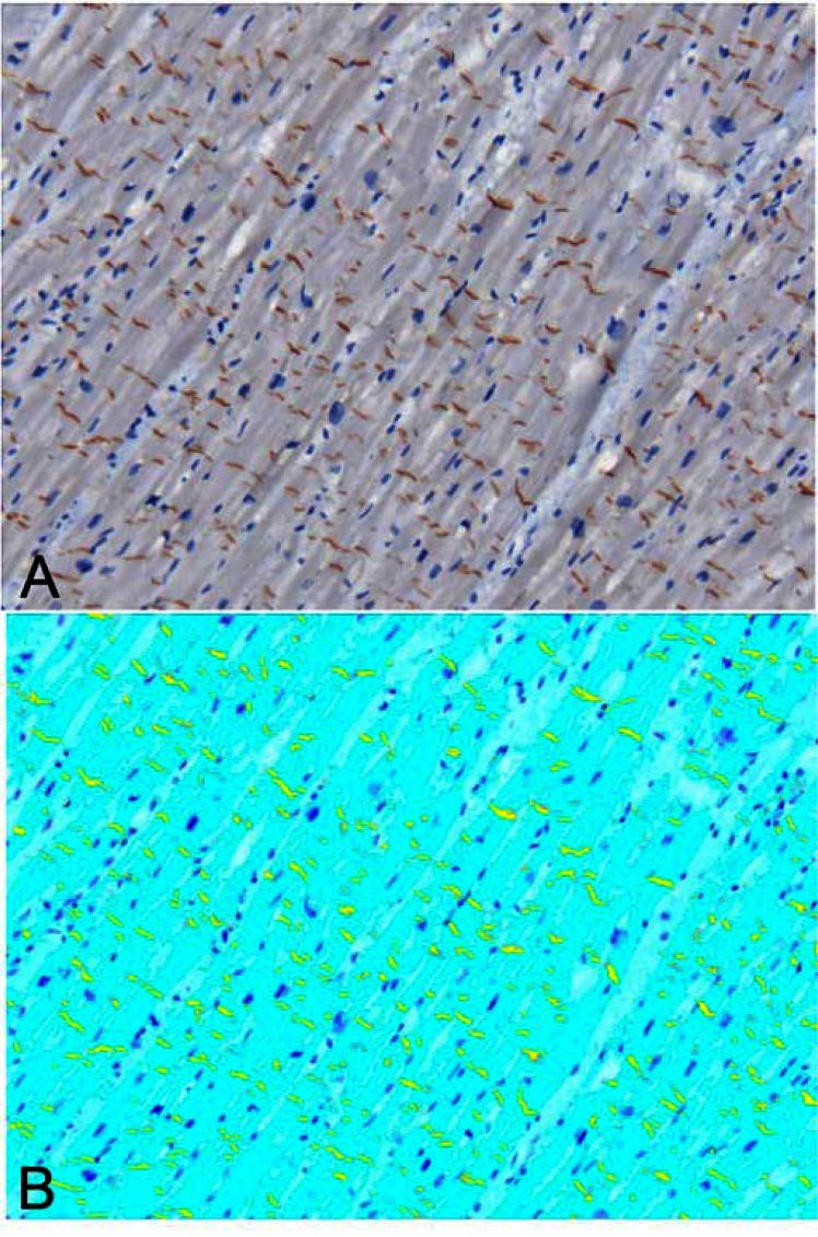
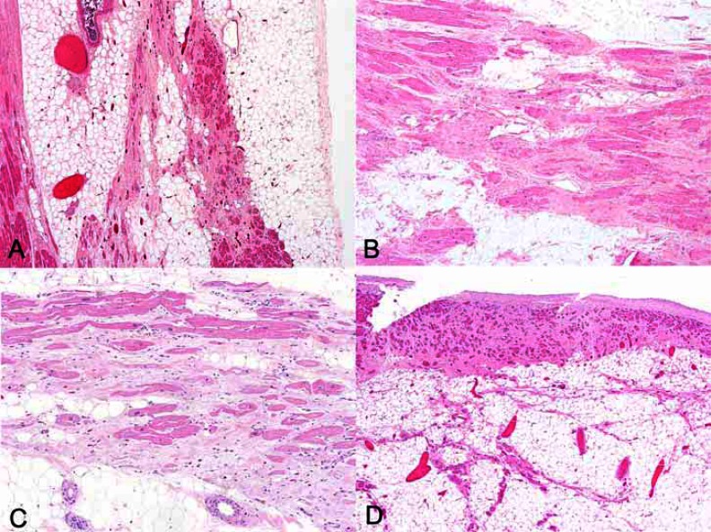
Methods and results: We studied 23 hearts from patients dying suddenly with ARVC. Control subject tissues were 21 hearts from people dying from non-cardiac causes (n=15), dilated cardiomyopathy (n=3) and coronary artery disease (n=3). Areas free of fibrofatty change or scarring were assessed on 50 sections from ARVC (24 left ventricle, 26 right ventricle) and 28 sections from controls. Immunohistochemical stains against plakoglobin, plakophilin, desmoplakin, connexin-43, and N-cadherin were applied and area expression analyzed by computerized morphometry. Desmin was stained as a control for fixation and similarly analyzed. The mean area of desmin expression was similar in controls and ARVC (86% vs. 85%, p=0.6). Plakoglobin expression was 4.9% ± 0.3% in controls, vs. 4.6% ± 0.3% in ARVC (p=0.3). Plakophilin staining was 4.8% ± 0.3% in controls vs. 4.4% ± 03% in ARVC (p=0.3). Desmoplakin staining was 3.4% in controls vs. 3.2 ± 0.2% in ARVC (p=0.6). There were no significant differences when staining was compared between right and left ventricles (all p > 0.1). For non-desmosomal proteins, the mean area of connexin-43 staining showed no significant difference by presence of disease.
Conclusions: The small and insignificant decrease in junction protein expression in ARVC suggests that immunohistochemistry is not a useful tool for the diagnosis.

 求助内容:
求助内容: 应助结果提醒方式:


