{"title":"光谱域 OCT 记录了假性囊样黄斑水肿在静脉注射曲安奈德后的消退情况。","authors":"Ravi K Murthy, Kakarla V Chalam","doi":"10.4137/oed.s3671","DOIUrl":null,"url":null,"abstract":"<p><p>Cystoid macular edema (CME) is an important cause of visual loss after cataract surgery. Treatment is usually with topical anti-inflammatory agents, with anti-vascular endothelial growth factor agents and steroids used intravitreally in resistant cases. Even though time-domain Stratus OCT can quantify the macular thickness, it cannot prognosticate visual outcomes due to the poor resolution of images, especially the outer segment-inner segment junction. Spectral-domain OCT (SD-OCT) by its ability to acquire large number of images in a short span of time provides high resolution cross-sectional images of the retina, which not only highlights the underlying pathological changes, but in addition can prognosticate visual recovery. We describe pre and post SD-OCT features of a case of refractory CME who was treated with intravitreal triamcinolone actetonide. </p>","PeriodicalId":74362,"journal":{"name":"Ophthalmology and eye diseases","volume":"2 ","pages":"1-4"},"PeriodicalIF":0.0000,"publicationDate":"2010-02-03","publicationTypes":"Journal Article","fieldsOfStudy":null,"isOpenAccess":false,"openAccessPdf":"https://www.ncbi.nlm.nih.gov/pmc/articles/PMC3661454/pdf/","citationCount":"0","resultStr":"{\"title\":\"Spectral Domain OCT Documented Resolution of Pseudophakic Cystoid Macular Edema after Intravitreal Triamcinolone.\",\"authors\":\"Ravi K Murthy, Kakarla V Chalam\",\"doi\":\"10.4137/oed.s3671\",\"DOIUrl\":null,\"url\":null,\"abstract\":\"<p><p>Cystoid macular edema (CME) is an important cause of visual loss after cataract surgery. Treatment is usually with topical anti-inflammatory agents, with anti-vascular endothelial growth factor agents and steroids used intravitreally in resistant cases. Even though time-domain Stratus OCT can quantify the macular thickness, it cannot prognosticate visual outcomes due to the poor resolution of images, especially the outer segment-inner segment junction. Spectral-domain OCT (SD-OCT) by its ability to acquire large number of images in a short span of time provides high resolution cross-sectional images of the retina, which not only highlights the underlying pathological changes, but in addition can prognosticate visual recovery. We describe pre and post SD-OCT features of a case of refractory CME who was treated with intravitreal triamcinolone actetonide. </p>\",\"PeriodicalId\":74362,\"journal\":{\"name\":\"Ophthalmology and eye diseases\",\"volume\":\"2 \",\"pages\":\"1-4\"},\"PeriodicalIF\":0.0000,\"publicationDate\":\"2010-02-03\",\"publicationTypes\":\"Journal Article\",\"fieldsOfStudy\":null,\"isOpenAccess\":false,\"openAccessPdf\":\"https://www.ncbi.nlm.nih.gov/pmc/articles/PMC3661454/pdf/\",\"citationCount\":\"0\",\"resultStr\":null,\"platform\":\"Semanticscholar\",\"paperid\":null,\"PeriodicalName\":\"Ophthalmology and eye diseases\",\"FirstCategoryId\":\"1085\",\"ListUrlMain\":\"https://doi.org/10.4137/oed.s3671\",\"RegionNum\":0,\"RegionCategory\":null,\"ArticlePicture\":[],\"TitleCN\":null,\"AbstractTextCN\":null,\"PMCID\":null,\"EPubDate\":\"2010/1/1 0:00:00\",\"PubModel\":\"Print\",\"JCR\":\"\",\"JCRName\":\"\",\"Score\":null,\"Total\":0}","platform":"Semanticscholar","paperid":null,"PeriodicalName":"Ophthalmology and eye diseases","FirstCategoryId":"1085","ListUrlMain":"https://doi.org/10.4137/oed.s3671","RegionNum":0,"RegionCategory":null,"ArticlePicture":[],"TitleCN":null,"AbstractTextCN":null,"PMCID":null,"EPubDate":"2010/1/1 0:00:00","PubModel":"Print","JCR":"","JCRName":"","Score":null,"Total":0}
引用次数: 0
摘要
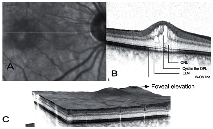
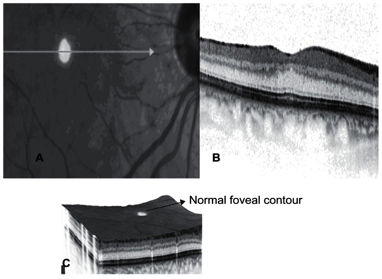
囊样黄斑水肿(CME)是白内障手术后视力下降的一个重要原因。治疗方法通常是局部使用抗炎药物,对于耐药的病例,可在玻璃体内使用抗血管内皮生长因子药物和类固醇。尽管时域斯特拉图 OCT 可以量化黄斑厚度,但由于图像分辨率较低,尤其是外节段与内节段交界处的图像,因此无法预测视觉结果。光谱域 OCT(SD-OCT)能在短时间内获取大量图像,提供高分辨率的视网膜横截面图像,不仅能突出显示潜在的病理变化,还能预示视力恢复情况。我们描述了一例难治性 CME 患者接受玻璃体内曲安奈德曲安奈德治疗前后的 SD-OCT 特征。
Spectral Domain OCT Documented Resolution of Pseudophakic Cystoid Macular Edema after Intravitreal Triamcinolone.
Cystoid macular edema (CME) is an important cause of visual loss after cataract surgery. Treatment is usually with topical anti-inflammatory agents, with anti-vascular endothelial growth factor agents and steroids used intravitreally in resistant cases. Even though time-domain Stratus OCT can quantify the macular thickness, it cannot prognosticate visual outcomes due to the poor resolution of images, especially the outer segment-inner segment junction. Spectral-domain OCT (SD-OCT) by its ability to acquire large number of images in a short span of time provides high resolution cross-sectional images of the retina, which not only highlights the underlying pathological changes, but in addition can prognosticate visual recovery. We describe pre and post SD-OCT features of a case of refractory CME who was treated with intravitreal triamcinolone actetonide.

 求助内容:
求助内容: 应助结果提醒方式:


