Gerard A Tarulli, Peter G Stanton, Kate L Loveland, Ewa Rajpert-De Meyts, Robert I McLachlan, Sarah J Meachem
{"title":"促性腺激素抑制和睾丸癌后男性支持细胞分化的研究。","authors":"Gerard A Tarulli, Peter G Stanton, Kate L Loveland, Ewa Rajpert-De Meyts, Robert I McLachlan, Sarah J Meachem","doi":"10.4161/spmg.24014","DOIUrl":null,"url":null,"abstract":"<p><p>It is widely held that the somatic cell population that is responsible for sperm development and output (Sertoli cells) is terminally differentiated and unmodifiable in adults. It is postulated, with little evidence, that Sertoli cells are not terminally differentiated in some phenotypes of infertility and testicular cancer. This study sought to compare markers of Sertoli cell differentiation in normospermic men, oligospermic men (undergoing gonadotropin suppression) and testicular carcinoma in situ (CIS) and seminoma samples. Confocal microscopy was used to assess the expression of markers of proliferation (PCNA and Ki67) and functional differentiation (androgen receptor). As additional markers of differentiation, the organization of Sertoli cell tight junction and associated proteins were assessed in specimens with carcinoma in situ. In normal men, Sertoli cells exhibited a differentiated phenotype (i.e., PCNA and Ki67 negative, androgen 40 receptor positive). However, after long-term gonadotropin suppression, 1.7 ± 0.6% of Sertoli cells exhibited PCNA reactivity associated with a diminished immunoreactivity in androgen receptor, suggesting an undifferentiated phenotype. Ki67-positive Sertoli cells were also observed. PCNA-positive Sertoli cells were never observed in tubules with carcinoma in situ, and only rarely observed adjacent to seminoma. Tight junction protein localization (claudin 11, JAM-A and ZO-1) was altered in CIS, with a reduction in JAM-A reactivity in Sertoli cells from tubules with CIS and the emergence of strong JAM-A reactivity in seminoma. These findings indicate that adult human Sertoli cells exhibit characteristics of an undifferentiated state in oligospermic men and patients with CIS and seminoma in the presence of germ cell neoplasia.</p>","PeriodicalId":22074,"journal":{"name":"Spermatogenesis","volume":"3 1","pages":"e24014"},"PeriodicalIF":0.0000,"publicationDate":"2013-01-01","publicationTypes":"Journal Article","fieldsOfStudy":null,"isOpenAccess":false,"openAccessPdf":"https://sci-hub-pdf.com/10.4161/spmg.24014","citationCount":"44","resultStr":"{\"title\":\"A survey of Sertoli cell differentiation in men after gonadotropin suppression and in testicular cancer.\",\"authors\":\"Gerard A Tarulli, Peter G Stanton, Kate L Loveland, Ewa Rajpert-De Meyts, Robert I McLachlan, Sarah J Meachem\",\"doi\":\"10.4161/spmg.24014\",\"DOIUrl\":null,\"url\":null,\"abstract\":\"<p><p>It is widely held that the somatic cell population that is responsible for sperm development and output (Sertoli cells) is terminally differentiated and unmodifiable in adults. It is postulated, with little evidence, that Sertoli cells are not terminally differentiated in some phenotypes of infertility and testicular cancer. This study sought to compare markers of Sertoli cell differentiation in normospermic men, oligospermic men (undergoing gonadotropin suppression) and testicular carcinoma in situ (CIS) and seminoma samples. Confocal microscopy was used to assess the expression of markers of proliferation (PCNA and Ki67) and functional differentiation (androgen receptor). As additional markers of differentiation, the organization of Sertoli cell tight junction and associated proteins were assessed in specimens with carcinoma in situ. In normal men, Sertoli cells exhibited a differentiated phenotype (i.e., PCNA and Ki67 negative, androgen 40 receptor positive). However, after long-term gonadotropin suppression, 1.7 ± 0.6% of Sertoli cells exhibited PCNA reactivity associated with a diminished immunoreactivity in androgen receptor, suggesting an undifferentiated phenotype. Ki67-positive Sertoli cells were also observed. PCNA-positive Sertoli cells were never observed in tubules with carcinoma in situ, and only rarely observed adjacent to seminoma. Tight junction protein localization (claudin 11, JAM-A and ZO-1) was altered in CIS, with a reduction in JAM-A reactivity in Sertoli cells from tubules with CIS and the emergence of strong JAM-A reactivity in seminoma. These findings indicate that adult human Sertoli cells exhibit characteristics of an undifferentiated state in oligospermic men and patients with CIS and seminoma in the presence of germ cell neoplasia.</p>\",\"PeriodicalId\":22074,\"journal\":{\"name\":\"Spermatogenesis\",\"volume\":\"3 1\",\"pages\":\"e24014\"},\"PeriodicalIF\":0.0000,\"publicationDate\":\"2013-01-01\",\"publicationTypes\":\"Journal Article\",\"fieldsOfStudy\":null,\"isOpenAccess\":false,\"openAccessPdf\":\"https://sci-hub-pdf.com/10.4161/spmg.24014\",\"citationCount\":\"44\",\"resultStr\":null,\"platform\":\"Semanticscholar\",\"paperid\":null,\"PeriodicalName\":\"Spermatogenesis\",\"FirstCategoryId\":\"1085\",\"ListUrlMain\":\"https://doi.org/10.4161/spmg.24014\",\"RegionNum\":0,\"RegionCategory\":null,\"ArticlePicture\":[],\"TitleCN\":null,\"AbstractTextCN\":null,\"PMCID\":null,\"EPubDate\":\"\",\"PubModel\":\"\",\"JCR\":\"\",\"JCRName\":\"\",\"Score\":null,\"Total\":0}","platform":"Semanticscholar","paperid":null,"PeriodicalName":"Spermatogenesis","FirstCategoryId":"1085","ListUrlMain":"https://doi.org/10.4161/spmg.24014","RegionNum":0,"RegionCategory":null,"ArticlePicture":[],"TitleCN":null,"AbstractTextCN":null,"PMCID":null,"EPubDate":"","PubModel":"","JCR":"","JCRName":"","Score":null,"Total":0}
A survey of Sertoli cell differentiation in men after gonadotropin suppression and in testicular cancer.
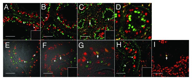
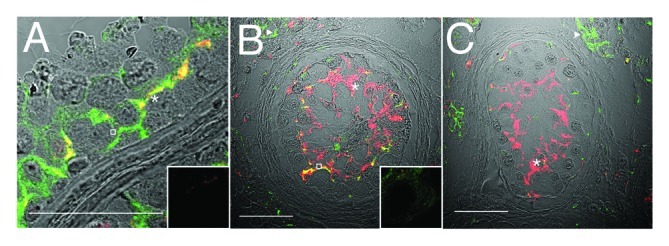
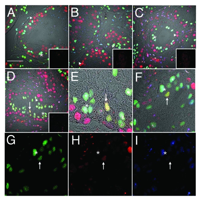
It is widely held that the somatic cell population that is responsible for sperm development and output (Sertoli cells) is terminally differentiated and unmodifiable in adults. It is postulated, with little evidence, that Sertoli cells are not terminally differentiated in some phenotypes of infertility and testicular cancer. This study sought to compare markers of Sertoli cell differentiation in normospermic men, oligospermic men (undergoing gonadotropin suppression) and testicular carcinoma in situ (CIS) and seminoma samples. Confocal microscopy was used to assess the expression of markers of proliferation (PCNA and Ki67) and functional differentiation (androgen receptor). As additional markers of differentiation, the organization of Sertoli cell tight junction and associated proteins were assessed in specimens with carcinoma in situ. In normal men, Sertoli cells exhibited a differentiated phenotype (i.e., PCNA and Ki67 negative, androgen 40 receptor positive). However, after long-term gonadotropin suppression, 1.7 ± 0.6% of Sertoli cells exhibited PCNA reactivity associated with a diminished immunoreactivity in androgen receptor, suggesting an undifferentiated phenotype. Ki67-positive Sertoli cells were also observed. PCNA-positive Sertoli cells were never observed in tubules with carcinoma in situ, and only rarely observed adjacent to seminoma. Tight junction protein localization (claudin 11, JAM-A and ZO-1) was altered in CIS, with a reduction in JAM-A reactivity in Sertoli cells from tubules with CIS and the emergence of strong JAM-A reactivity in seminoma. These findings indicate that adult human Sertoli cells exhibit characteristics of an undifferentiated state in oligospermic men and patients with CIS and seminoma in the presence of germ cell neoplasia.

 求助内容:
求助内容: 应助结果提醒方式:


