{"title":"继发性CML伴骨髓纤维化的髓外细胞危象。","authors":"Jai Hyang Go, Joowon Park","doi":"10.5045/kjh.2012.47.4.243","DOIUrl":null,"url":null,"abstract":"which permits unrestricted non-commercial use, distribution, and reproduction in any medium, provided the original work is properly cited. A 54-year-old man was referred to our hospital with right flank pain. Three years ago, he was diagnosed with gastric mucosa-associated lymphoid tissue (MALT) lymphoma and successfully treated with radiotherapy. CBC showed a WBC count of 24. Bone marrow (BM) examination showed granulocytic and megakaryocytic proliferation with moderate dysplastic megakaryopoiesis (A; H&E stain, ×200), and diffuse reticulin fibrosis (B; reticulin stain, ×400). Primary myelofibrosis was the first diagnostic consideration after BM study. Chromosomal analysis, however, showed t(9;22)(q34;q11.2), indicating CML. Concurrent abdomen computerized tomography revealed enlarged inguinal lymph nodes. Inguinal lymph node biopsy showed diffuse infiltration of immature cells (C; H&E stain, ×400), which were positive for myeloperoxidase (D). BCR/ABL1 rearrangement was demonstrated by fluorescence in-situ hybridization analysis, and a diagnosis of granulocytic sarcoma (GS) was made. Accompanying extramedullary myeloid tumor, CML was classified as blastic phase. Secondary CML with a simultaneous manifestation of GS is rare. Combining morphological and molecular-cytogenetic approaches can help detect the coexistence of both neoplasms, especially in CML cases with fewer typical morphologic features.","PeriodicalId":23001,"journal":{"name":"The Korean Journal of Hematology","volume":"47 4","pages":"243"},"PeriodicalIF":0.0000,"publicationDate":"2012-12-01","publicationTypes":"Journal Article","fieldsOfStudy":null,"isOpenAccess":false,"openAccessPdf":"https://sci-hub-pdf.com/10.5045/kjh.2012.47.4.243","citationCount":"3","resultStr":"{\"title\":\"Extramedullary blast crisis of secondary CML accompanying marrow fibrosis.\",\"authors\":\"Jai Hyang Go, Joowon Park\",\"doi\":\"10.5045/kjh.2012.47.4.243\",\"DOIUrl\":null,\"url\":null,\"abstract\":\"which permits unrestricted non-commercial use, distribution, and reproduction in any medium, provided the original work is properly cited. A 54-year-old man was referred to our hospital with right flank pain. Three years ago, he was diagnosed with gastric mucosa-associated lymphoid tissue (MALT) lymphoma and successfully treated with radiotherapy. CBC showed a WBC count of 24. Bone marrow (BM) examination showed granulocytic and megakaryocytic proliferation with moderate dysplastic megakaryopoiesis (A; H&E stain, ×200), and diffuse reticulin fibrosis (B; reticulin stain, ×400). Primary myelofibrosis was the first diagnostic consideration after BM study. Chromosomal analysis, however, showed t(9;22)(q34;q11.2), indicating CML. Concurrent abdomen computerized tomography revealed enlarged inguinal lymph nodes. Inguinal lymph node biopsy showed diffuse infiltration of immature cells (C; H&E stain, ×400), which were positive for myeloperoxidase (D). BCR/ABL1 rearrangement was demonstrated by fluorescence in-situ hybridization analysis, and a diagnosis of granulocytic sarcoma (GS) was made. Accompanying extramedullary myeloid tumor, CML was classified as blastic phase. Secondary CML with a simultaneous manifestation of GS is rare. Combining morphological and molecular-cytogenetic approaches can help detect the coexistence of both neoplasms, especially in CML cases with fewer typical morphologic features.\",\"PeriodicalId\":23001,\"journal\":{\"name\":\"The Korean Journal of Hematology\",\"volume\":\"47 4\",\"pages\":\"243\"},\"PeriodicalIF\":0.0000,\"publicationDate\":\"2012-12-01\",\"publicationTypes\":\"Journal Article\",\"fieldsOfStudy\":null,\"isOpenAccess\":false,\"openAccessPdf\":\"https://sci-hub-pdf.com/10.5045/kjh.2012.47.4.243\",\"citationCount\":\"3\",\"resultStr\":null,\"platform\":\"Semanticscholar\",\"paperid\":null,\"PeriodicalName\":\"The Korean Journal of Hematology\",\"FirstCategoryId\":\"1085\",\"ListUrlMain\":\"https://doi.org/10.5045/kjh.2012.47.4.243\",\"RegionNum\":0,\"RegionCategory\":null,\"ArticlePicture\":[],\"TitleCN\":null,\"AbstractTextCN\":null,\"PMCID\":null,\"EPubDate\":\"2012/12/24 0:00:00\",\"PubModel\":\"Epub\",\"JCR\":\"\",\"JCRName\":\"\",\"Score\":null,\"Total\":0}","platform":"Semanticscholar","paperid":null,"PeriodicalName":"The Korean Journal of Hematology","FirstCategoryId":"1085","ListUrlMain":"https://doi.org/10.5045/kjh.2012.47.4.243","RegionNum":0,"RegionCategory":null,"ArticlePicture":[],"TitleCN":null,"AbstractTextCN":null,"PMCID":null,"EPubDate":"2012/12/24 0:00:00","PubModel":"Epub","JCR":"","JCRName":"","Score":null,"Total":0}
Extramedullary blast crisis of secondary CML accompanying marrow fibrosis.
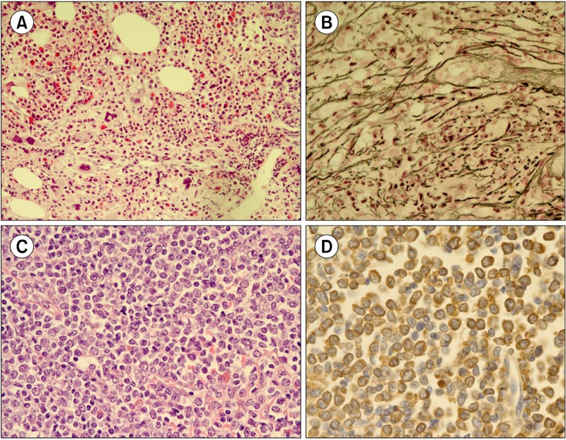
which permits unrestricted non-commercial use, distribution, and reproduction in any medium, provided the original work is properly cited. A 54-year-old man was referred to our hospital with right flank pain. Three years ago, he was diagnosed with gastric mucosa-associated lymphoid tissue (MALT) lymphoma and successfully treated with radiotherapy. CBC showed a WBC count of 24. Bone marrow (BM) examination showed granulocytic and megakaryocytic proliferation with moderate dysplastic megakaryopoiesis (A; H&E stain, ×200), and diffuse reticulin fibrosis (B; reticulin stain, ×400). Primary myelofibrosis was the first diagnostic consideration after BM study. Chromosomal analysis, however, showed t(9;22)(q34;q11.2), indicating CML. Concurrent abdomen computerized tomography revealed enlarged inguinal lymph nodes. Inguinal lymph node biopsy showed diffuse infiltration of immature cells (C; H&E stain, ×400), which were positive for myeloperoxidase (D). BCR/ABL1 rearrangement was demonstrated by fluorescence in-situ hybridization analysis, and a diagnosis of granulocytic sarcoma (GS) was made. Accompanying extramedullary myeloid tumor, CML was classified as blastic phase. Secondary CML with a simultaneous manifestation of GS is rare. Combining morphological and molecular-cytogenetic approaches can help detect the coexistence of both neoplasms, especially in CML cases with fewer typical morphologic features.

 求助内容:
求助内容: 应助结果提醒方式:


