Carlos Eduardo Oliveira Dos Santos, Daniele Malaman, César Vivian Lopes, Júlio Carlos Pereira-Lima, Artur Adolfo Parada
{"title":"数字彩色内镜在结肠小病变诊断中的应用。","authors":"Carlos Eduardo Oliveira Dos Santos, Daniele Malaman, César Vivian Lopes, Júlio Carlos Pereira-Lima, Artur Adolfo Parada","doi":"10.1155/2012/279521","DOIUrl":null,"url":null,"abstract":"<p><p>Introduction. To compare the accuracy of digital and real-time chromoendoscopy for the differential diagnosis of diminutive (<5 mm) neoplastic and nonneoplastic colorectal lesions. Materials and Methods. This is a prospective randomized study comparing the Fujinon intelligent color enhancement (FICE) system (65 patients/95 lesions) and indigo carmine (69 patients/120 lesions) in the analysis of capillary meshwork and pit pattern, respectively. All lesions were less than 5 mm in diameter, and magnification was used in both groups. Histopathology was the gold standard examination. Results. Of 215 colorectal lesions, 153 (71.2%) were adenomas, and 62 were hyperplastic polyps (28.8%). Morphological analysis revealed 132 (61.4%) superficial lesions, with 7 (3.3%) depressed lesions, and 83 (38.6%) protruding lesions. Vascular meshwork analysis using FICE and magnification resulted in 91.7% sensitivity, 95.7% specificity, and 92.6% accuracy in differentiating neoplastic from nonneoplastic lesions. Pit pattern analysis with indigo carmine and magnification showed 96.5% sensitivity, 88.2% specificity, and 94.2% accuracy for the same purpose. Conclusion. Both magnifying virtual chromoendoscopy and indigo carmine chromoendoscopy showed high accuracy in the histopathological diagnosis of colorectal lesions less than 5 mm in diameter.</p>","PeriodicalId":11288,"journal":{"name":"Diagnostic and Therapeutic Endoscopy","volume":"2012 ","pages":"279521"},"PeriodicalIF":0.0000,"publicationDate":"2012-01-01","publicationTypes":"Journal Article","fieldsOfStudy":null,"isOpenAccess":false,"openAccessPdf":"https://sci-hub-pdf.com/10.1155/2012/279521","citationCount":"19","resultStr":"{\"title\":\"Digital chromoendoscopy for diagnosis of diminutive colorectal lesions.\",\"authors\":\"Carlos Eduardo Oliveira Dos Santos, Daniele Malaman, César Vivian Lopes, Júlio Carlos Pereira-Lima, Artur Adolfo Parada\",\"doi\":\"10.1155/2012/279521\",\"DOIUrl\":null,\"url\":null,\"abstract\":\"<p><p>Introduction. To compare the accuracy of digital and real-time chromoendoscopy for the differential diagnosis of diminutive (<5 mm) neoplastic and nonneoplastic colorectal lesions. Materials and Methods. This is a prospective randomized study comparing the Fujinon intelligent color enhancement (FICE) system (65 patients/95 lesions) and indigo carmine (69 patients/120 lesions) in the analysis of capillary meshwork and pit pattern, respectively. All lesions were less than 5 mm in diameter, and magnification was used in both groups. Histopathology was the gold standard examination. Results. Of 215 colorectal lesions, 153 (71.2%) were adenomas, and 62 were hyperplastic polyps (28.8%). Morphological analysis revealed 132 (61.4%) superficial lesions, with 7 (3.3%) depressed lesions, and 83 (38.6%) protruding lesions. Vascular meshwork analysis using FICE and magnification resulted in 91.7% sensitivity, 95.7% specificity, and 92.6% accuracy in differentiating neoplastic from nonneoplastic lesions. Pit pattern analysis with indigo carmine and magnification showed 96.5% sensitivity, 88.2% specificity, and 94.2% accuracy for the same purpose. Conclusion. Both magnifying virtual chromoendoscopy and indigo carmine chromoendoscopy showed high accuracy in the histopathological diagnosis of colorectal lesions less than 5 mm in diameter.</p>\",\"PeriodicalId\":11288,\"journal\":{\"name\":\"Diagnostic and Therapeutic Endoscopy\",\"volume\":\"2012 \",\"pages\":\"279521\"},\"PeriodicalIF\":0.0000,\"publicationDate\":\"2012-01-01\",\"publicationTypes\":\"Journal Article\",\"fieldsOfStudy\":null,\"isOpenAccess\":false,\"openAccessPdf\":\"https://sci-hub-pdf.com/10.1155/2012/279521\",\"citationCount\":\"19\",\"resultStr\":null,\"platform\":\"Semanticscholar\",\"paperid\":null,\"PeriodicalName\":\"Diagnostic and Therapeutic Endoscopy\",\"FirstCategoryId\":\"1085\",\"ListUrlMain\":\"https://doi.org/10.1155/2012/279521\",\"RegionNum\":0,\"RegionCategory\":null,\"ArticlePicture\":[],\"TitleCN\":null,\"AbstractTextCN\":null,\"PMCID\":null,\"EPubDate\":\"2012/10/3 0:00:00\",\"PubModel\":\"Epub\",\"JCR\":\"\",\"JCRName\":\"\",\"Score\":null,\"Total\":0}","platform":"Semanticscholar","paperid":null,"PeriodicalName":"Diagnostic and Therapeutic Endoscopy","FirstCategoryId":"1085","ListUrlMain":"https://doi.org/10.1155/2012/279521","RegionNum":0,"RegionCategory":null,"ArticlePicture":[],"TitleCN":null,"AbstractTextCN":null,"PMCID":null,"EPubDate":"2012/10/3 0:00:00","PubModel":"Epub","JCR":"","JCRName":"","Score":null,"Total":0}
Digital chromoendoscopy for diagnosis of diminutive colorectal lesions.
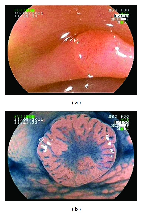
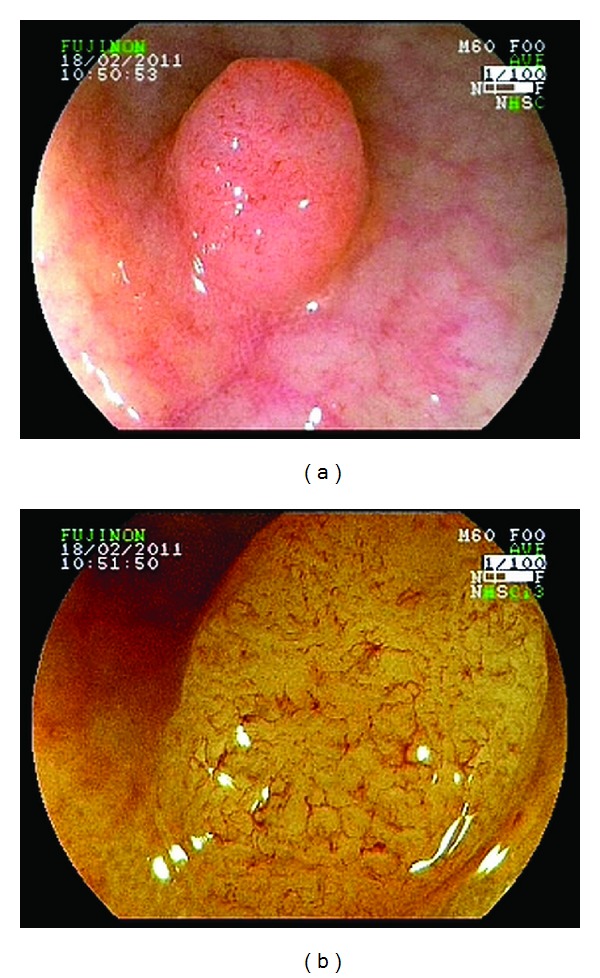
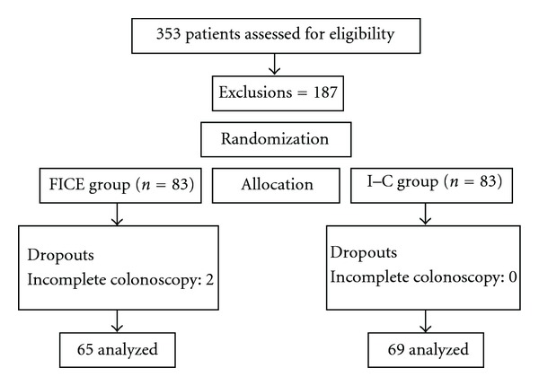
Introduction. To compare the accuracy of digital and real-time chromoendoscopy for the differential diagnosis of diminutive (<5 mm) neoplastic and nonneoplastic colorectal lesions. Materials and Methods. This is a prospective randomized study comparing the Fujinon intelligent color enhancement (FICE) system (65 patients/95 lesions) and indigo carmine (69 patients/120 lesions) in the analysis of capillary meshwork and pit pattern, respectively. All lesions were less than 5 mm in diameter, and magnification was used in both groups. Histopathology was the gold standard examination. Results. Of 215 colorectal lesions, 153 (71.2%) were adenomas, and 62 were hyperplastic polyps (28.8%). Morphological analysis revealed 132 (61.4%) superficial lesions, with 7 (3.3%) depressed lesions, and 83 (38.6%) protruding lesions. Vascular meshwork analysis using FICE and magnification resulted in 91.7% sensitivity, 95.7% specificity, and 92.6% accuracy in differentiating neoplastic from nonneoplastic lesions. Pit pattern analysis with indigo carmine and magnification showed 96.5% sensitivity, 88.2% specificity, and 94.2% accuracy for the same purpose. Conclusion. Both magnifying virtual chromoendoscopy and indigo carmine chromoendoscopy showed high accuracy in the histopathological diagnosis of colorectal lesions less than 5 mm in diameter.

 求助内容:
求助内容: 应助结果提醒方式:


