Zhenghui Wang, Ke Zhang, Karen L Wooley, John-Stephen Taylor
{"title":"通过 PNA-DNA 链置换激活探针成像活细胞中的 mRNA 表达。","authors":"Zhenghui Wang, Ke Zhang, Karen L Wooley, John-Stephen Taylor","doi":"10.1155/2012/962652","DOIUrl":null,"url":null,"abstract":"<p><p>Probes for monitoring mRNA expression in vivo are of great interest for the study of biological and biomedical problems, but progress has been hampered by poor signal to noise and effective means for delivering the probes into live cells. Herein we report a PNA·DNA strand displacement-activated fluorescent probe that can image the expression of iNOS (inducible nitric oxide synthase) mRNA, a marker of inflammation. The probe consists of a fluorescein labeled antisense PNA annealed to a shorter DABCYL(plus)-labeled DNA which quenches the fluorescence, but when the quencher strand is displaced by the target mRNA the fluorescence is restored. DNA was used for the quencher strand to facilitate electrostatic binding of the otherwise netural PNA strand to a cationic shell crosslinked knedel-like (cSCK) nanoparticle which can deliver the PNA·DNA duplex probe into cells with less toxicity and greater efficiency than other transfection agents. RAW 264.7 mouse macrophage cells transfected with the iNOS PNA·DNA probe via the cSCK showed a 16 to 54-fold increase in average fluorescence per cell upon iNOS stimulation. The increase was 4 to 7-fold higher than that for a non-complementary probe, thereby validating the ability of a PNA·DNA strand displacement-activated probe to image mRNA expression in vivo.</p>","PeriodicalId":16575,"journal":{"name":"Journal of Nucleic Acids","volume":"2012 ","pages":"962652"},"PeriodicalIF":1.3000,"publicationDate":"2012-01-01","publicationTypes":"Journal Article","fieldsOfStudy":null,"isOpenAccess":false,"openAccessPdf":"https://www.ncbi.nlm.nih.gov/pmc/articles/PMC3463960/pdf/","citationCount":"0","resultStr":"{\"title\":\"Imaging mRNA Expression in Live Cells via PNA·DNA Strand Displacement-Activated Probes.\",\"authors\":\"Zhenghui Wang, Ke Zhang, Karen L Wooley, John-Stephen Taylor\",\"doi\":\"10.1155/2012/962652\",\"DOIUrl\":null,\"url\":null,\"abstract\":\"<p><p>Probes for monitoring mRNA expression in vivo are of great interest for the study of biological and biomedical problems, but progress has been hampered by poor signal to noise and effective means for delivering the probes into live cells. Herein we report a PNA·DNA strand displacement-activated fluorescent probe that can image the expression of iNOS (inducible nitric oxide synthase) mRNA, a marker of inflammation. The probe consists of a fluorescein labeled antisense PNA annealed to a shorter DABCYL(plus)-labeled DNA which quenches the fluorescence, but when the quencher strand is displaced by the target mRNA the fluorescence is restored. DNA was used for the quencher strand to facilitate electrostatic binding of the otherwise netural PNA strand to a cationic shell crosslinked knedel-like (cSCK) nanoparticle which can deliver the PNA·DNA duplex probe into cells with less toxicity and greater efficiency than other transfection agents. RAW 264.7 mouse macrophage cells transfected with the iNOS PNA·DNA probe via the cSCK showed a 16 to 54-fold increase in average fluorescence per cell upon iNOS stimulation. The increase was 4 to 7-fold higher than that for a non-complementary probe, thereby validating the ability of a PNA·DNA strand displacement-activated probe to image mRNA expression in vivo.</p>\",\"PeriodicalId\":16575,\"journal\":{\"name\":\"Journal of Nucleic Acids\",\"volume\":\"2012 \",\"pages\":\"962652\"},\"PeriodicalIF\":1.3000,\"publicationDate\":\"2012-01-01\",\"publicationTypes\":\"Journal Article\",\"fieldsOfStudy\":null,\"isOpenAccess\":false,\"openAccessPdf\":\"https://www.ncbi.nlm.nih.gov/pmc/articles/PMC3463960/pdf/\",\"citationCount\":\"0\",\"resultStr\":null,\"platform\":\"Semanticscholar\",\"paperid\":null,\"PeriodicalName\":\"Journal of Nucleic Acids\",\"FirstCategoryId\":\"1085\",\"ListUrlMain\":\"https://doi.org/10.1155/2012/962652\",\"RegionNum\":0,\"RegionCategory\":null,\"ArticlePicture\":[],\"TitleCN\":null,\"AbstractTextCN\":null,\"PMCID\":null,\"EPubDate\":\"2012/9/26 0:00:00\",\"PubModel\":\"Epub\",\"JCR\":\"Q4\",\"JCRName\":\"BIOCHEMISTRY & MOLECULAR BIOLOGY\",\"Score\":null,\"Total\":0}","platform":"Semanticscholar","paperid":null,"PeriodicalName":"Journal of Nucleic Acids","FirstCategoryId":"1085","ListUrlMain":"https://doi.org/10.1155/2012/962652","RegionNum":0,"RegionCategory":null,"ArticlePicture":[],"TitleCN":null,"AbstractTextCN":null,"PMCID":null,"EPubDate":"2012/9/26 0:00:00","PubModel":"Epub","JCR":"Q4","JCRName":"BIOCHEMISTRY & MOLECULAR BIOLOGY","Score":null,"Total":0}
Imaging mRNA Expression in Live Cells via PNA·DNA Strand Displacement-Activated Probes.
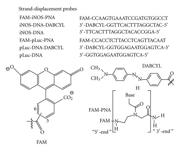
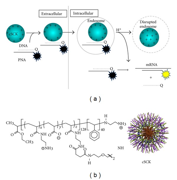
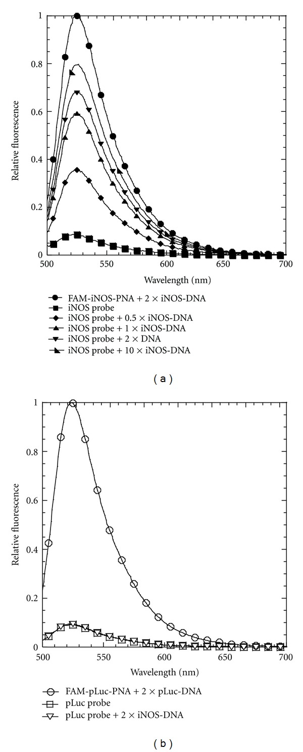
Probes for monitoring mRNA expression in vivo are of great interest for the study of biological and biomedical problems, but progress has been hampered by poor signal to noise and effective means for delivering the probes into live cells. Herein we report a PNA·DNA strand displacement-activated fluorescent probe that can image the expression of iNOS (inducible nitric oxide synthase) mRNA, a marker of inflammation. The probe consists of a fluorescein labeled antisense PNA annealed to a shorter DABCYL(plus)-labeled DNA which quenches the fluorescence, but when the quencher strand is displaced by the target mRNA the fluorescence is restored. DNA was used for the quencher strand to facilitate electrostatic binding of the otherwise netural PNA strand to a cationic shell crosslinked knedel-like (cSCK) nanoparticle which can deliver the PNA·DNA duplex probe into cells with less toxicity and greater efficiency than other transfection agents. RAW 264.7 mouse macrophage cells transfected with the iNOS PNA·DNA probe via the cSCK showed a 16 to 54-fold increase in average fluorescence per cell upon iNOS stimulation. The increase was 4 to 7-fold higher than that for a non-complementary probe, thereby validating the ability of a PNA·DNA strand displacement-activated probe to image mRNA expression in vivo.

 求助内容:
求助内容: 应助结果提醒方式:


