Frits A Rangel, Thomas J J Maal, Stefaan J Bergé, Anne Marie Kuijpers-Jagtman
{"title":"锥形束计算机断层扫描中数字牙科模型的集成。","authors":"Frits A Rangel, Thomas J J Maal, Stefaan J Bergé, Anne Marie Kuijpers-Jagtman","doi":"10.5402/2012/949086","DOIUrl":null,"url":null,"abstract":"<p><p>Cone-beam computed tomography (CBCT) is widely used in maxillofacial surgery. The CBCT image of the dental arches, however, is of insufficient quality to use in digital planning of orthognathic surgery. Several authors have described methods to integrate digital dental casts into CBCT scans, but all reported methods have drawbacks. The aim of this feasibility study is to present a new simplified method to integrate digital dental casts into CBCT scans. In a patient scheduled for orthognathic surgery, titanium markers were glued to the gingiva. Next, a CBCT scan and dental impressions were made. During the impression-taking procedure, the titanium markers were transferred to the impression. The impressions were scanned, and all CBCT datasets were exported in DICOM format. The two datasets were matched, and the dentition derived from the scanned impressions was transferred to the CBCT of the patient. After matching the two datasets, the average distance between the corresponding markers was 0.1 mm. This novel method allows for the integration of digital dental casts into CBCT scans, overcoming problems such as unwanted extra radiation exposure, distortion of soft tissues due to the use of bite jigs, and time-consuming digital data handling.</p>","PeriodicalId":89396,"journal":{"name":"ISRN dentistry","volume":"2012 ","pages":"949086"},"PeriodicalIF":0.0000,"publicationDate":"2012-01-01","publicationTypes":"Journal Article","fieldsOfStudy":null,"isOpenAccess":false,"openAccessPdf":"https://www.ncbi.nlm.nih.gov/pmc/articles/PMC3461639/pdf/","citationCount":"26","resultStr":"{\"title\":\"Integration of digital dental casts in cone-beam computed tomography scans.\",\"authors\":\"Frits A Rangel, Thomas J J Maal, Stefaan J Bergé, Anne Marie Kuijpers-Jagtman\",\"doi\":\"10.5402/2012/949086\",\"DOIUrl\":null,\"url\":null,\"abstract\":\"<p><p>Cone-beam computed tomography (CBCT) is widely used in maxillofacial surgery. The CBCT image of the dental arches, however, is of insufficient quality to use in digital planning of orthognathic surgery. Several authors have described methods to integrate digital dental casts into CBCT scans, but all reported methods have drawbacks. The aim of this feasibility study is to present a new simplified method to integrate digital dental casts into CBCT scans. In a patient scheduled for orthognathic surgery, titanium markers were glued to the gingiva. Next, a CBCT scan and dental impressions were made. During the impression-taking procedure, the titanium markers were transferred to the impression. The impressions were scanned, and all CBCT datasets were exported in DICOM format. The two datasets were matched, and the dentition derived from the scanned impressions was transferred to the CBCT of the patient. After matching the two datasets, the average distance between the corresponding markers was 0.1 mm. This novel method allows for the integration of digital dental casts into CBCT scans, overcoming problems such as unwanted extra radiation exposure, distortion of soft tissues due to the use of bite jigs, and time-consuming digital data handling.</p>\",\"PeriodicalId\":89396,\"journal\":{\"name\":\"ISRN dentistry\",\"volume\":\"2012 \",\"pages\":\"949086\"},\"PeriodicalIF\":0.0000,\"publicationDate\":\"2012-01-01\",\"publicationTypes\":\"Journal Article\",\"fieldsOfStudy\":null,\"isOpenAccess\":false,\"openAccessPdf\":\"https://www.ncbi.nlm.nih.gov/pmc/articles/PMC3461639/pdf/\",\"citationCount\":\"26\",\"resultStr\":null,\"platform\":\"Semanticscholar\",\"paperid\":null,\"PeriodicalName\":\"ISRN dentistry\",\"FirstCategoryId\":\"1085\",\"ListUrlMain\":\"https://doi.org/10.5402/2012/949086\",\"RegionNum\":0,\"RegionCategory\":null,\"ArticlePicture\":[],\"TitleCN\":null,\"AbstractTextCN\":null,\"PMCID\":null,\"EPubDate\":\"2012/9/23 0:00:00\",\"PubModel\":\"Epub\",\"JCR\":\"\",\"JCRName\":\"\",\"Score\":null,\"Total\":0}","platform":"Semanticscholar","paperid":null,"PeriodicalName":"ISRN dentistry","FirstCategoryId":"1085","ListUrlMain":"https://doi.org/10.5402/2012/949086","RegionNum":0,"RegionCategory":null,"ArticlePicture":[],"TitleCN":null,"AbstractTextCN":null,"PMCID":null,"EPubDate":"2012/9/23 0:00:00","PubModel":"Epub","JCR":"","JCRName":"","Score":null,"Total":0}
Integration of digital dental casts in cone-beam computed tomography scans.
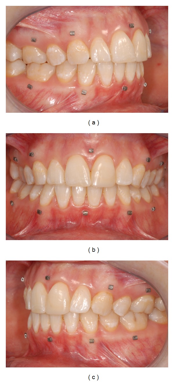
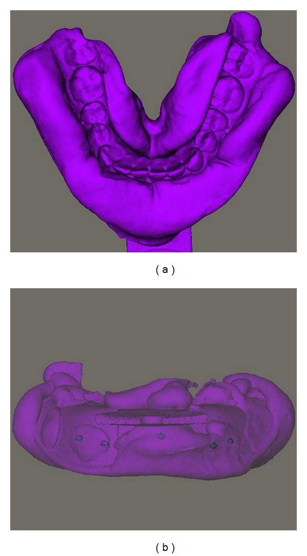
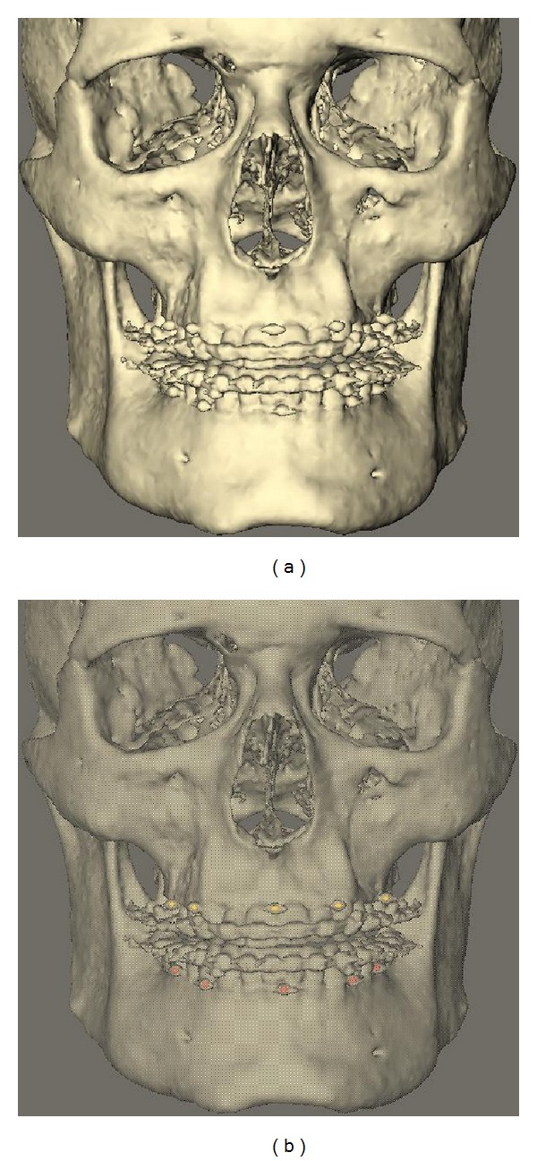
Cone-beam computed tomography (CBCT) is widely used in maxillofacial surgery. The CBCT image of the dental arches, however, is of insufficient quality to use in digital planning of orthognathic surgery. Several authors have described methods to integrate digital dental casts into CBCT scans, but all reported methods have drawbacks. The aim of this feasibility study is to present a new simplified method to integrate digital dental casts into CBCT scans. In a patient scheduled for orthognathic surgery, titanium markers were glued to the gingiva. Next, a CBCT scan and dental impressions were made. During the impression-taking procedure, the titanium markers were transferred to the impression. The impressions were scanned, and all CBCT datasets were exported in DICOM format. The two datasets were matched, and the dentition derived from the scanned impressions was transferred to the CBCT of the patient. After matching the two datasets, the average distance between the corresponding markers was 0.1 mm. This novel method allows for the integration of digital dental casts into CBCT scans, overcoming problems such as unwanted extra radiation exposure, distortion of soft tissues due to the use of bite jigs, and time-consuming digital data handling.

 求助内容:
求助内容: 应助结果提醒方式:


