{"title":"泰国人肺静脉引流的解剖变异:多探头CT研究。","authors":"Y Wannasopha, N Oilmungmool, J Euathrongchit","doi":"10.2349/biij.8.1.e4","DOIUrl":null,"url":null,"abstract":"<p><strong>Objective: </strong>To evaluate the patterns of pulmonary venous drainage into the left atrium and to determine the frequency of each variant of pulmonary venous anatomy.</p><p><strong>Materials and methods: </strong>After institutional review board approval (No. 09JUL011148), 300 studies of thoracic multidetector computed tomography were retrospectively reviewed for the anatomical features of the pulmonary vein and its drainage pattern into the left atrium. The percentage of each pattern was calculated.</p><p><strong>Results: </strong>The anatomy of pulmonary venous drainage in 300 patients (150 male and 150 female, mean age 60.16 years) showed some variation. In the right pulmonary vein, the most common drainage pattern was two ostia (90.33%), followed by three to five ostia (6.33%) and a single ostium (3.33%). There were one or two separate middle lobe vein ostia in groups of more than two openings. On the left side, there were two patterns; a single venous ostium (59%) was much more common than two ostia (41%). In both right and left pulmonary veins, there were five cases (2 male, 3 female) that had a single pulmonary venous ostium, bilaterally. However, there were only 17 cases (5.67%), out of 300 enrolled in this study, that had bilateral pulmonary venous ostial variations.</p><p><strong>Conclusion: </strong>A classification system to succinctly describe pulmonary venous drainage patterns was developed. In left-sided drainage, a single left pulmonary ostium was the most common variation. The right-sided venous drainage varied more in both number and pattern than those of the left side; nevertheless, bilateral pulmonary venous ostial variation was not frequently found.</p>","PeriodicalId":89331,"journal":{"name":"Biomedical imaging and intervention journal","volume":"8 1","pages":"e4"},"PeriodicalIF":0.0000,"publicationDate":"2012-01-01","publicationTypes":"Journal Article","fieldsOfStudy":null,"isOpenAccess":false,"openAccessPdf":"https://sci-hub-pdf.com/10.2349/biij.8.1.e4","citationCount":"0","resultStr":"{\"title\":\"Anatomical variations of pulmonary venous drainage in Thai people: multidetector CT study.\",\"authors\":\"Y Wannasopha, N Oilmungmool, J Euathrongchit\",\"doi\":\"10.2349/biij.8.1.e4\",\"DOIUrl\":null,\"url\":null,\"abstract\":\"<p><strong>Objective: </strong>To evaluate the patterns of pulmonary venous drainage into the left atrium and to determine the frequency of each variant of pulmonary venous anatomy.</p><p><strong>Materials and methods: </strong>After institutional review board approval (No. 09JUL011148), 300 studies of thoracic multidetector computed tomography were retrospectively reviewed for the anatomical features of the pulmonary vein and its drainage pattern into the left atrium. The percentage of each pattern was calculated.</p><p><strong>Results: </strong>The anatomy of pulmonary venous drainage in 300 patients (150 male and 150 female, mean age 60.16 years) showed some variation. In the right pulmonary vein, the most common drainage pattern was two ostia (90.33%), followed by three to five ostia (6.33%) and a single ostium (3.33%). There were one or two separate middle lobe vein ostia in groups of more than two openings. On the left side, there were two patterns; a single venous ostium (59%) was much more common than two ostia (41%). In both right and left pulmonary veins, there were five cases (2 male, 3 female) that had a single pulmonary venous ostium, bilaterally. However, there were only 17 cases (5.67%), out of 300 enrolled in this study, that had bilateral pulmonary venous ostial variations.</p><p><strong>Conclusion: </strong>A classification system to succinctly describe pulmonary venous drainage patterns was developed. In left-sided drainage, a single left pulmonary ostium was the most common variation. The right-sided venous drainage varied more in both number and pattern than those of the left side; nevertheless, bilateral pulmonary venous ostial variation was not frequently found.</p>\",\"PeriodicalId\":89331,\"journal\":{\"name\":\"Biomedical imaging and intervention journal\",\"volume\":\"8 1\",\"pages\":\"e4\"},\"PeriodicalIF\":0.0000,\"publicationDate\":\"2012-01-01\",\"publicationTypes\":\"Journal Article\",\"fieldsOfStudy\":null,\"isOpenAccess\":false,\"openAccessPdf\":\"https://sci-hub-pdf.com/10.2349/biij.8.1.e4\",\"citationCount\":\"0\",\"resultStr\":null,\"platform\":\"Semanticscholar\",\"paperid\":null,\"PeriodicalName\":\"Biomedical imaging and intervention journal\",\"FirstCategoryId\":\"1085\",\"ListUrlMain\":\"https://doi.org/10.2349/biij.8.1.e4\",\"RegionNum\":0,\"RegionCategory\":null,\"ArticlePicture\":[],\"TitleCN\":null,\"AbstractTextCN\":null,\"PMCID\":null,\"EPubDate\":\"\",\"PubModel\":\"\",\"JCR\":\"\",\"JCRName\":\"\",\"Score\":null,\"Total\":0}","platform":"Semanticscholar","paperid":null,"PeriodicalName":"Biomedical imaging and intervention journal","FirstCategoryId":"1085","ListUrlMain":"https://doi.org/10.2349/biij.8.1.e4","RegionNum":0,"RegionCategory":null,"ArticlePicture":[],"TitleCN":null,"AbstractTextCN":null,"PMCID":null,"EPubDate":"","PubModel":"","JCR":"","JCRName":"","Score":null,"Total":0}
Anatomical variations of pulmonary venous drainage in Thai people: multidetector CT study.
Objective: To evaluate the patterns of pulmonary venous drainage into the left atrium and to determine the frequency of each variant of pulmonary venous anatomy.
Materials and methods: After institutional review board approval (No. 09JUL011148), 300 studies of thoracic multidetector computed tomography were retrospectively reviewed for the anatomical features of the pulmonary vein and its drainage pattern into the left atrium. The percentage of each pattern was calculated.
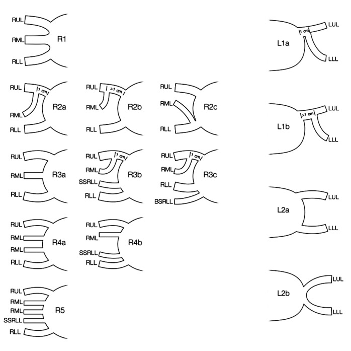
Results: The anatomy of pulmonary venous drainage in 300 patients (150 male and 150 female, mean age 60.16 years) showed some variation. In the right pulmonary vein, the most common drainage pattern was two ostia (90.33%), followed by three to five ostia (6.33%) and a single ostium (3.33%). There were one or two separate middle lobe vein ostia in groups of more than two openings. On the left side, there were two patterns; a single venous ostium (59%) was much more common than two ostia (41%). In both right and left pulmonary veins, there were five cases (2 male, 3 female) that had a single pulmonary venous ostium, bilaterally. However, there were only 17 cases (5.67%), out of 300 enrolled in this study, that had bilateral pulmonary venous ostial variations.
Conclusion: A classification system to succinctly describe pulmonary venous drainage patterns was developed. In left-sided drainage, a single left pulmonary ostium was the most common variation. The right-sided venous drainage varied more in both number and pattern than those of the left side; nevertheless, bilateral pulmonary venous ostial variation was not frequently found.

 求助内容:
求助内容: 应助结果提醒方式:


