Vincent J D Krouwer, Liesbeth H P Hekking, Miriam Langelaar-Makkinje, Elsa Regan-Klapisz, Jan Andries Post
{"title":"内皮细胞衰老与细胞间连接中断和单层通透性增加有关。","authors":"Vincent J D Krouwer, Liesbeth H P Hekking, Miriam Langelaar-Makkinje, Elsa Regan-Klapisz, Jan Andries Post","doi":"10.1186/2045-824X-4-12","DOIUrl":null,"url":null,"abstract":"<p><strong>Unlabelled: </strong></p><p><strong>Background: </strong>Cellular senescence is associated with cellular dysfunction and has been shown to occur in vivo in age-related cardiovascular diseases such as atherosclerosis. Atherogenesis is accompanied by intimal accumulation of LDL and increased extravasation of monocytes towards accumulated and oxidized LDL, suggesting an affected barrier function of vascular endothelial cells. Our objective was to study the effect of cellular senescence on the barrier function of non-senescent endothelial cells.</p><p><strong>Methods: </strong>Human umbilical vein endothelial cells were cultured until senescence. Senescent cells were compared with non-senescent cells and with co-cultures of non-senescent and senescent cells. Adherens junctions and tight junctions were studied. To assess the barrier function of various monolayers, assays to measure permeability for Lucifer Yellow (LY) and horseradish peroxidase (PO) were performed.</p><p><strong>Results: </strong>The barrier function of monolayers comprising of senescent cells was compromised and coincided with a change in the distribution of junction proteins and a down-regulation of occludin and claudin-5 expression. Furthermore, a decreased expression of occludin and claudin-5 was observed in co-cultures of non-senescent and senescent cells, not only between senescent cells but also along the entire periphery of non-senescent cells lining a senescent cell.</p><p><strong>Conclusions: </strong>Our findings show that the presence of senescent endothelial cells in a non-senescent monolayer disrupts tight junction morphology of surrounding young cells and increases the permeability of the monolayer for LY and PO.</p>","PeriodicalId":23948,"journal":{"name":"Vascular Cell","volume":"4 1","pages":"12"},"PeriodicalIF":0.0000,"publicationDate":"2012-08-28","publicationTypes":"Journal Article","fieldsOfStudy":null,"isOpenAccess":false,"openAccessPdf":"https://sci-hub-pdf.com/10.1186/2045-824X-4-12","citationCount":"99","resultStr":"{\"title\":\"Endothelial cell senescence is associated with disrupted cell-cell junctions and increased monolayer permeability.\",\"authors\":\"Vincent J D Krouwer, Liesbeth H P Hekking, Miriam Langelaar-Makkinje, Elsa Regan-Klapisz, Jan Andries Post\",\"doi\":\"10.1186/2045-824X-4-12\",\"DOIUrl\":null,\"url\":null,\"abstract\":\"<p><strong>Unlabelled: </strong></p><p><strong>Background: </strong>Cellular senescence is associated with cellular dysfunction and has been shown to occur in vivo in age-related cardiovascular diseases such as atherosclerosis. Atherogenesis is accompanied by intimal accumulation of LDL and increased extravasation of monocytes towards accumulated and oxidized LDL, suggesting an affected barrier function of vascular endothelial cells. Our objective was to study the effect of cellular senescence on the barrier function of non-senescent endothelial cells.</p><p><strong>Methods: </strong>Human umbilical vein endothelial cells were cultured until senescence. Senescent cells were compared with non-senescent cells and with co-cultures of non-senescent and senescent cells. Adherens junctions and tight junctions were studied. To assess the barrier function of various monolayers, assays to measure permeability for Lucifer Yellow (LY) and horseradish peroxidase (PO) were performed.</p><p><strong>Results: </strong>The barrier function of monolayers comprising of senescent cells was compromised and coincided with a change in the distribution of junction proteins and a down-regulation of occludin and claudin-5 expression. Furthermore, a decreased expression of occludin and claudin-5 was observed in co-cultures of non-senescent and senescent cells, not only between senescent cells but also along the entire periphery of non-senescent cells lining a senescent cell.</p><p><strong>Conclusions: </strong>Our findings show that the presence of senescent endothelial cells in a non-senescent monolayer disrupts tight junction morphology of surrounding young cells and increases the permeability of the monolayer for LY and PO.</p>\",\"PeriodicalId\":23948,\"journal\":{\"name\":\"Vascular Cell\",\"volume\":\"4 1\",\"pages\":\"12\"},\"PeriodicalIF\":0.0000,\"publicationDate\":\"2012-08-28\",\"publicationTypes\":\"Journal Article\",\"fieldsOfStudy\":null,\"isOpenAccess\":false,\"openAccessPdf\":\"https://sci-hub-pdf.com/10.1186/2045-824X-4-12\",\"citationCount\":\"99\",\"resultStr\":null,\"platform\":\"Semanticscholar\",\"paperid\":null,\"PeriodicalName\":\"Vascular Cell\",\"FirstCategoryId\":\"1085\",\"ListUrlMain\":\"https://doi.org/10.1186/2045-824X-4-12\",\"RegionNum\":0,\"RegionCategory\":null,\"ArticlePicture\":[],\"TitleCN\":null,\"AbstractTextCN\":null,\"PMCID\":null,\"EPubDate\":\"\",\"PubModel\":\"\",\"JCR\":\"Q4\",\"JCRName\":\"Neuroscience\",\"Score\":null,\"Total\":0}","platform":"Semanticscholar","paperid":null,"PeriodicalName":"Vascular Cell","FirstCategoryId":"1085","ListUrlMain":"https://doi.org/10.1186/2045-824X-4-12","RegionNum":0,"RegionCategory":null,"ArticlePicture":[],"TitleCN":null,"AbstractTextCN":null,"PMCID":null,"EPubDate":"","PubModel":"","JCR":"Q4","JCRName":"Neuroscience","Score":null,"Total":0}
Endothelial cell senescence is associated with disrupted cell-cell junctions and increased monolayer permeability.
Unlabelled:
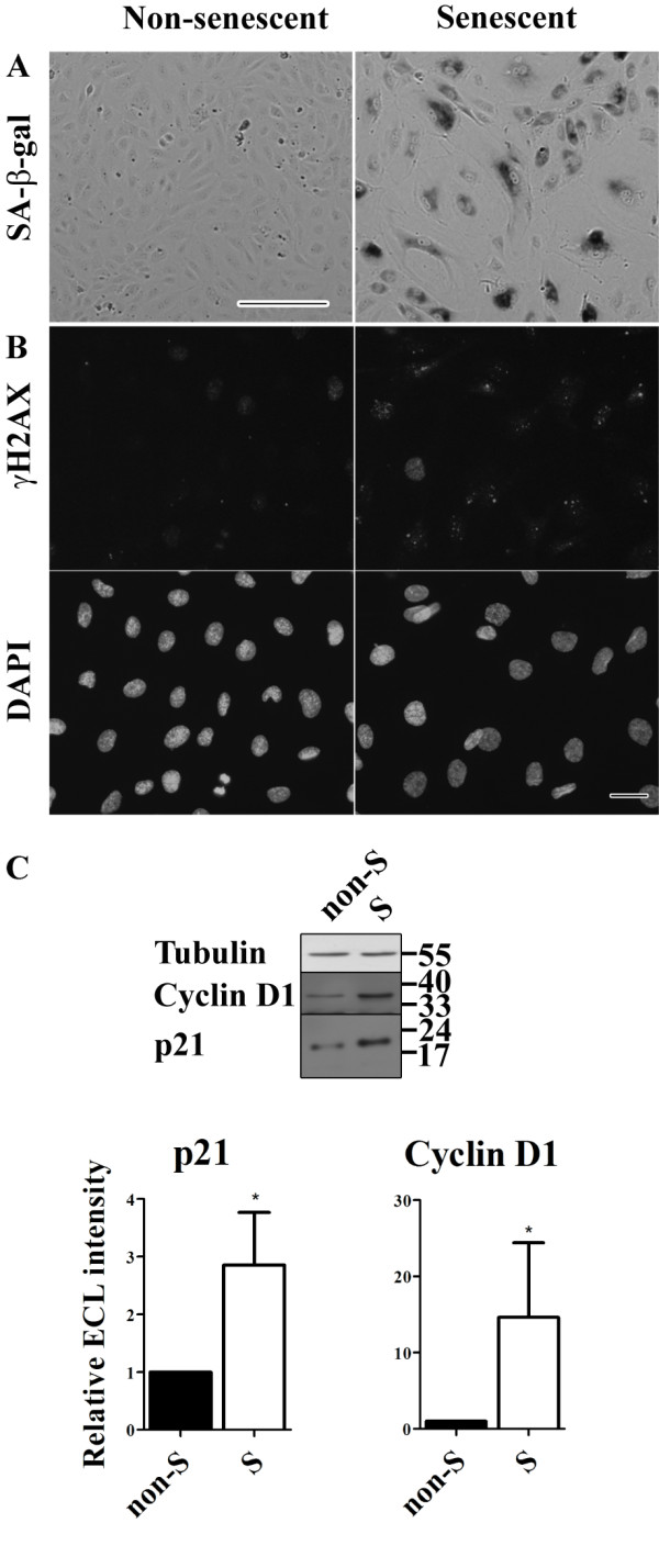
Background: Cellular senescence is associated with cellular dysfunction and has been shown to occur in vivo in age-related cardiovascular diseases such as atherosclerosis. Atherogenesis is accompanied by intimal accumulation of LDL and increased extravasation of monocytes towards accumulated and oxidized LDL, suggesting an affected barrier function of vascular endothelial cells. Our objective was to study the effect of cellular senescence on the barrier function of non-senescent endothelial cells.
Methods: Human umbilical vein endothelial cells were cultured until senescence. Senescent cells were compared with non-senescent cells and with co-cultures of non-senescent and senescent cells. Adherens junctions and tight junctions were studied. To assess the barrier function of various monolayers, assays to measure permeability for Lucifer Yellow (LY) and horseradish peroxidase (PO) were performed.
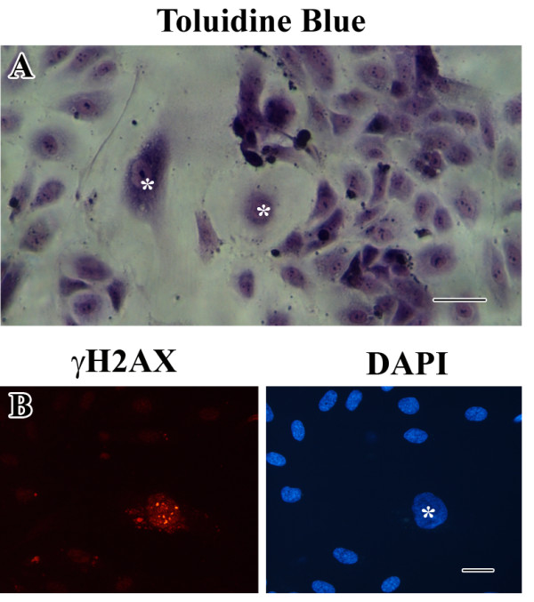
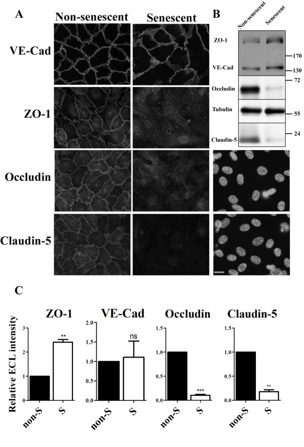
Results: The barrier function of monolayers comprising of senescent cells was compromised and coincided with a change in the distribution of junction proteins and a down-regulation of occludin and claudin-5 expression. Furthermore, a decreased expression of occludin and claudin-5 was observed in co-cultures of non-senescent and senescent cells, not only between senescent cells but also along the entire periphery of non-senescent cells lining a senescent cell.
Conclusions: Our findings show that the presence of senescent endothelial cells in a non-senescent monolayer disrupts tight junction morphology of surrounding young cells and increases the permeability of the monolayer for LY and PO.

 求助内容:
求助内容: 应助结果提醒方式:


