{"title":"三种两栖动物肝脏结构的比较组织学研究。","authors":"Hideo Akiyoshi, Asuka M Inoue","doi":"10.1186/1476-5926-11-2","DOIUrl":null,"url":null,"abstract":"<p><strong>Unlabelled: </strong></p><p><strong>Background: </strong>This report presents a detailed description of hepatic architecture in 46 amphibian livers by light microscopy, and extensively discusses the phylogenetic viewpoint.</p><p><strong>Results: </strong>The 46 amphibian livers showed a variety of histological features, but anurans were the same as in mammalian livers. The hepatocyte-sinusoidal structures of the amphibian livers were classified into three different types: (I) several-cell-thick plate type, (II) two-cell-thick plate type, and (III) one-cell-thick plate type, depending on the percentage extension of sinusoidal areas per unit area, measured by morphometry. Hematopoietic tissue structures were observed in the connective tissue of both the perihepatic subcapsular regions and portal triads in the order Caudata and Gymnophiona, but were not observed in the order Anura (except for the genus Bombina and Xenopus). As phylogenetic relationships are branched from urodeles to anurans, the parenchyma arrangement progressed from the combined several- and two-cell-thick plate type to one-cell-thick plate type as seen in the mammalian liver type. In contrast, hematopoietic tissue structures were exactly the opposite and did not involve anurans.</p><p><strong>Conclusions: </strong>This study is the first to investigate amphibian livers phylogenically, and their architectural differences are shown in the route of hepatic ontogenesis. In this process, parenchymal arrangement formation is acquired phylogenically. The occurrence of hematopoietic cells may be related with the development of the systemic immune system in the spleen and bone marrow.</p>","PeriodicalId":84474,"journal":{"name":"Comparative hepatology","volume":"11 1","pages":"2"},"PeriodicalIF":0.0000,"publicationDate":"2012-08-20","publicationTypes":"Journal Article","fieldsOfStudy":null,"isOpenAccess":false,"openAccessPdf":"https://www.ncbi.nlm.nih.gov/pmc/articles/PMC3517316/pdf/","citationCount":"0","resultStr":"{\"title\":\"Comparative histological study of hepatic architecture in the three orders amphibian livers.\",\"authors\":\"Hideo Akiyoshi, Asuka M Inoue\",\"doi\":\"10.1186/1476-5926-11-2\",\"DOIUrl\":null,\"url\":null,\"abstract\":\"<p><strong>Unlabelled: </strong></p><p><strong>Background: </strong>This report presents a detailed description of hepatic architecture in 46 amphibian livers by light microscopy, and extensively discusses the phylogenetic viewpoint.</p><p><strong>Results: </strong>The 46 amphibian livers showed a variety of histological features, but anurans were the same as in mammalian livers. The hepatocyte-sinusoidal structures of the amphibian livers were classified into three different types: (I) several-cell-thick plate type, (II) two-cell-thick plate type, and (III) one-cell-thick plate type, depending on the percentage extension of sinusoidal areas per unit area, measured by morphometry. Hematopoietic tissue structures were observed in the connective tissue of both the perihepatic subcapsular regions and portal triads in the order Caudata and Gymnophiona, but were not observed in the order Anura (except for the genus Bombina and Xenopus). As phylogenetic relationships are branched from urodeles to anurans, the parenchyma arrangement progressed from the combined several- and two-cell-thick plate type to one-cell-thick plate type as seen in the mammalian liver type. In contrast, hematopoietic tissue structures were exactly the opposite and did not involve anurans.</p><p><strong>Conclusions: </strong>This study is the first to investigate amphibian livers phylogenically, and their architectural differences are shown in the route of hepatic ontogenesis. In this process, parenchymal arrangement formation is acquired phylogenically. The occurrence of hematopoietic cells may be related with the development of the systemic immune system in the spleen and bone marrow.</p>\",\"PeriodicalId\":84474,\"journal\":{\"name\":\"Comparative hepatology\",\"volume\":\"11 1\",\"pages\":\"2\"},\"PeriodicalIF\":0.0000,\"publicationDate\":\"2012-08-20\",\"publicationTypes\":\"Journal Article\",\"fieldsOfStudy\":null,\"isOpenAccess\":false,\"openAccessPdf\":\"https://www.ncbi.nlm.nih.gov/pmc/articles/PMC3517316/pdf/\",\"citationCount\":\"0\",\"resultStr\":null,\"platform\":\"Semanticscholar\",\"paperid\":null,\"PeriodicalName\":\"Comparative hepatology\",\"FirstCategoryId\":\"1085\",\"ListUrlMain\":\"https://doi.org/10.1186/1476-5926-11-2\",\"RegionNum\":0,\"RegionCategory\":null,\"ArticlePicture\":[],\"TitleCN\":null,\"AbstractTextCN\":null,\"PMCID\":null,\"EPubDate\":\"\",\"PubModel\":\"\",\"JCR\":\"\",\"JCRName\":\"\",\"Score\":null,\"Total\":0}","platform":"Semanticscholar","paperid":null,"PeriodicalName":"Comparative hepatology","FirstCategoryId":"1085","ListUrlMain":"https://doi.org/10.1186/1476-5926-11-2","RegionNum":0,"RegionCategory":null,"ArticlePicture":[],"TitleCN":null,"AbstractTextCN":null,"PMCID":null,"EPubDate":"","PubModel":"","JCR":"","JCRName":"","Score":null,"Total":0}
Comparative histological study of hepatic architecture in the three orders amphibian livers.
Unlabelled:
Background: This report presents a detailed description of hepatic architecture in 46 amphibian livers by light microscopy, and extensively discusses the phylogenetic viewpoint.
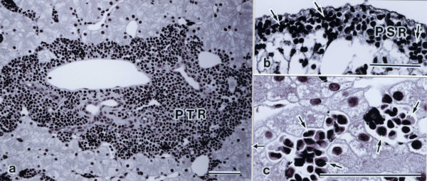
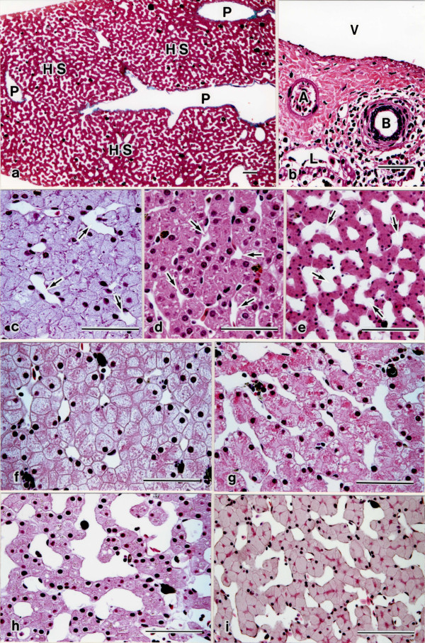
Results: The 46 amphibian livers showed a variety of histological features, but anurans were the same as in mammalian livers. The hepatocyte-sinusoidal structures of the amphibian livers were classified into three different types: (I) several-cell-thick plate type, (II) two-cell-thick plate type, and (III) one-cell-thick plate type, depending on the percentage extension of sinusoidal areas per unit area, measured by morphometry. Hematopoietic tissue structures were observed in the connective tissue of both the perihepatic subcapsular regions and portal triads in the order Caudata and Gymnophiona, but were not observed in the order Anura (except for the genus Bombina and Xenopus). As phylogenetic relationships are branched from urodeles to anurans, the parenchyma arrangement progressed from the combined several- and two-cell-thick plate type to one-cell-thick plate type as seen in the mammalian liver type. In contrast, hematopoietic tissue structures were exactly the opposite and did not involve anurans.
Conclusions: This study is the first to investigate amphibian livers phylogenically, and their architectural differences are shown in the route of hepatic ontogenesis. In this process, parenchymal arrangement formation is acquired phylogenically. The occurrence of hematopoietic cells may be related with the development of the systemic immune system in the spleen and bone marrow.

 求助内容:
求助内容: 应助结果提醒方式:


