{"title":"丙型肝炎病毒核心蛋白基因型3的脂滴结合。","authors":"Guan Qiang, Ravi Jhaveri","doi":"10.5402/2012/176728","DOIUrl":null,"url":null,"abstract":"<p><p>Background. Hepatitis C virus (HCV) genotype 3 is known to cause steatosis (fatty liver) that is more frequent and severe than other genotypes. We previously identified sequence elements within genotype 3 HCV Core domain 3 that were sufficient for lipid accumulation. Aims. We examined various genotype 3 Core domains for lipid droplet localization and compared the lipid droplet binding regions of domain 2 with a genotype 1 isolate. Methods. We generated HCV Core domain constructs fused with green fluorescent protein and performed immunofluorescence to visualize lipid droplets. Results. Constructs containing HCV Core domain 2 are appropriately localized to lipid droplets with varying degrees of efficiency. When compared to genotype 1, there are polymorphisms within domain 2 that do not appear to alter lipid droplet localization. Conclusions. In summary, the differences in a steatosis-associated HCV Core genotype 3 isolate do not appear to involve altered lipid droplet localization.</p>","PeriodicalId":89397,"journal":{"name":"ISRN gastroenterology","volume":"2012 ","pages":"176728"},"PeriodicalIF":0.0000,"publicationDate":"2012-01-01","publicationTypes":"Journal Article","fieldsOfStudy":null,"isOpenAccess":false,"openAccessPdf":"https://sci-hub-pdf.com/10.5402/2012/176728","citationCount":"8","resultStr":"{\"title\":\"Lipid droplet binding of hepatitis C virus core protein genotype 3.\",\"authors\":\"Guan Qiang, Ravi Jhaveri\",\"doi\":\"10.5402/2012/176728\",\"DOIUrl\":null,\"url\":null,\"abstract\":\"<p><p>Background. Hepatitis C virus (HCV) genotype 3 is known to cause steatosis (fatty liver) that is more frequent and severe than other genotypes. We previously identified sequence elements within genotype 3 HCV Core domain 3 that were sufficient for lipid accumulation. Aims. We examined various genotype 3 Core domains for lipid droplet localization and compared the lipid droplet binding regions of domain 2 with a genotype 1 isolate. Methods. We generated HCV Core domain constructs fused with green fluorescent protein and performed immunofluorescence to visualize lipid droplets. Results. Constructs containing HCV Core domain 2 are appropriately localized to lipid droplets with varying degrees of efficiency. When compared to genotype 1, there are polymorphisms within domain 2 that do not appear to alter lipid droplet localization. Conclusions. In summary, the differences in a steatosis-associated HCV Core genotype 3 isolate do not appear to involve altered lipid droplet localization.</p>\",\"PeriodicalId\":89397,\"journal\":{\"name\":\"ISRN gastroenterology\",\"volume\":\"2012 \",\"pages\":\"176728\"},\"PeriodicalIF\":0.0000,\"publicationDate\":\"2012-01-01\",\"publicationTypes\":\"Journal Article\",\"fieldsOfStudy\":null,\"isOpenAccess\":false,\"openAccessPdf\":\"https://sci-hub-pdf.com/10.5402/2012/176728\",\"citationCount\":\"8\",\"resultStr\":null,\"platform\":\"Semanticscholar\",\"paperid\":null,\"PeriodicalName\":\"ISRN gastroenterology\",\"FirstCategoryId\":\"1085\",\"ListUrlMain\":\"https://doi.org/10.5402/2012/176728\",\"RegionNum\":0,\"RegionCategory\":null,\"ArticlePicture\":[],\"TitleCN\":null,\"AbstractTextCN\":null,\"PMCID\":null,\"EPubDate\":\"2012/7/11 0:00:00\",\"PubModel\":\"Epub\",\"JCR\":\"\",\"JCRName\":\"\",\"Score\":null,\"Total\":0}","platform":"Semanticscholar","paperid":null,"PeriodicalName":"ISRN gastroenterology","FirstCategoryId":"1085","ListUrlMain":"https://doi.org/10.5402/2012/176728","RegionNum":0,"RegionCategory":null,"ArticlePicture":[],"TitleCN":null,"AbstractTextCN":null,"PMCID":null,"EPubDate":"2012/7/11 0:00:00","PubModel":"Epub","JCR":"","JCRName":"","Score":null,"Total":0}
Lipid droplet binding of hepatitis C virus core protein genotype 3.
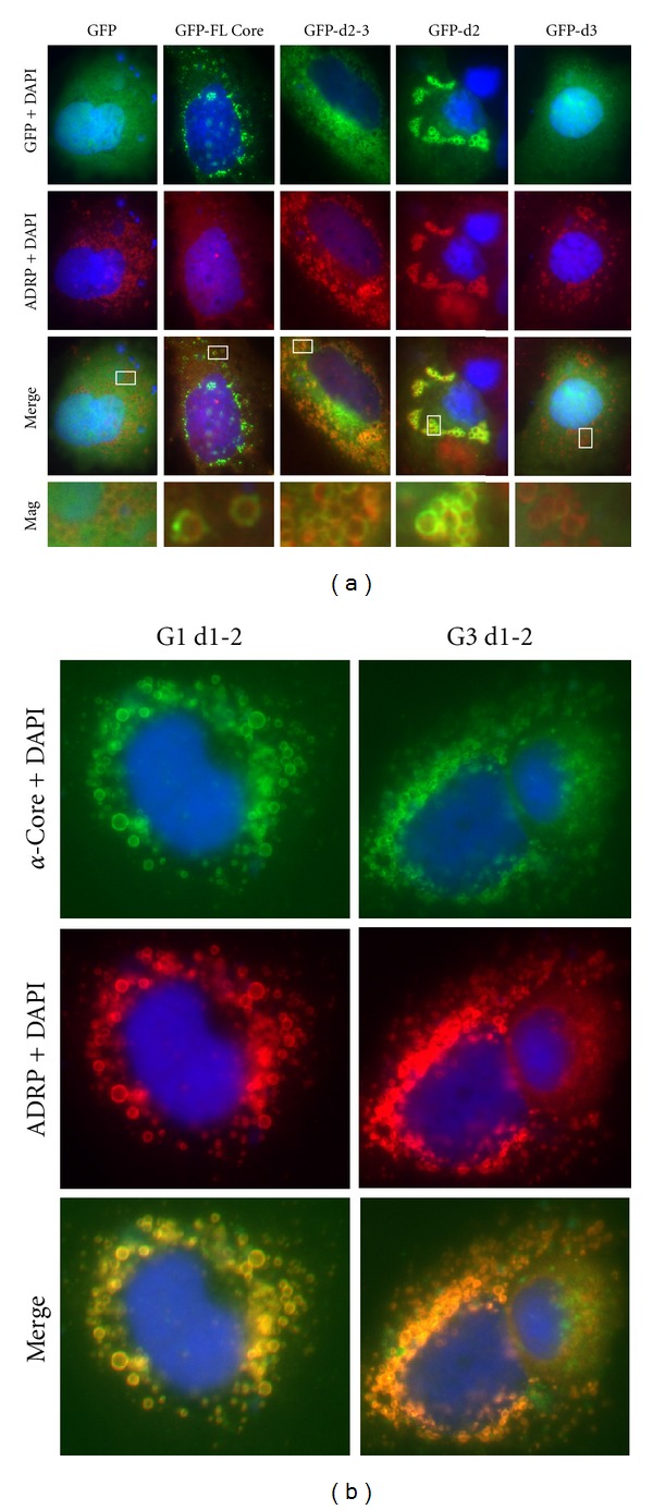
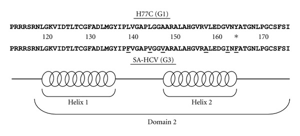
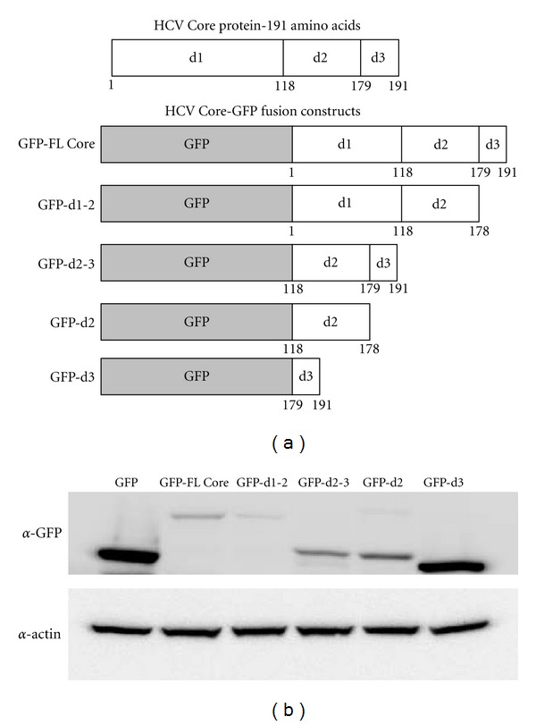
Background. Hepatitis C virus (HCV) genotype 3 is known to cause steatosis (fatty liver) that is more frequent and severe than other genotypes. We previously identified sequence elements within genotype 3 HCV Core domain 3 that were sufficient for lipid accumulation. Aims. We examined various genotype 3 Core domains for lipid droplet localization and compared the lipid droplet binding regions of domain 2 with a genotype 1 isolate. Methods. We generated HCV Core domain constructs fused with green fluorescent protein and performed immunofluorescence to visualize lipid droplets. Results. Constructs containing HCV Core domain 2 are appropriately localized to lipid droplets with varying degrees of efficiency. When compared to genotype 1, there are polymorphisms within domain 2 that do not appear to alter lipid droplet localization. Conclusions. In summary, the differences in a steatosis-associated HCV Core genotype 3 isolate do not appear to involve altered lipid droplet localization.

 求助内容:
求助内容: 应助结果提醒方式:


