Anne-Frédérique Manichon, Brigitte Bancel, Marion Durieux-Millon, Christian Ducerf, Jean-Yves Mabrut, Marie-Annick Lepogam, Agnès Rode
{"title":"肝细胞腺瘤:26例超声和MRI增强评价及病理和表型分型的相关性","authors":"Anne-Frédérique Manichon, Brigitte Bancel, Marion Durieux-Millon, Christian Ducerf, Jean-Yves Mabrut, Marie-Annick Lepogam, Agnès Rode","doi":"10.1155/2012/418745","DOIUrl":null,"url":null,"abstract":"<p><p>Purpose. To review the contrast-enhanced ultrasonographic (CEUS) and magnetic resonance (MR) imaging findings in 25 patients with 26 hepatocellular adenomas (HCAs) and to compare imaging features with histopathologic results from resected specimen considering the new immunophenotypical classification. Material and Methods. Two abdominal radiologists reviewed retrospectively CEUS cineloops and MR images in 26 HCA. All pathological specimens were reviewed and classified into four subgroups (steatotic or HNF 1α mutated, inflammatory, atypical or β-catenin mutated, and unspecified). Inflammatory infiltrates were scored, steatosis, and telangiectasia semiquantitatively evaluated. Results. CEUS and MRI features are well correlated: among the 16 inflammatory HCA, 7/16 presented typical imaging features: hypersignal T2, strong arterial enhancement with a centripetal filling, persistent on delayed phase. 6 HCA were classified as steatotic with typical imaging features: a drop out signal, slight arterial enhancement, vanishing on late phase. Four HCA were classified as atypical with an HCC developed in one. Five lesions displayed important steatosis (>50%) without belonging to the HNF1α group. Conclusion. In half cases, inflammatory HCA have specific imaging features well correlated with the amount of telangiectasia and inflammatory infiltrates. An HCA with important amount of steatosis noticed on chemical shift images does not always belong to the HNF1α group.</p>","PeriodicalId":77165,"journal":{"name":"HPB surgery : a world journal of hepatic, pancreatic and biliary surgery","volume":"2012 ","pages":"418745"},"PeriodicalIF":0.0000,"publicationDate":"2012-01-01","publicationTypes":"Journal Article","fieldsOfStudy":null,"isOpenAccess":false,"openAccessPdf":"https://sci-hub-pdf.com/10.1155/2012/418745","citationCount":"32","resultStr":"{\"title\":\"Hepatocellular adenoma: evaluation with contrast-enhanced ultrasound and MRI and correlation with pathologic and phenotypic classification in 26 lesions.\",\"authors\":\"Anne-Frédérique Manichon, Brigitte Bancel, Marion Durieux-Millon, Christian Ducerf, Jean-Yves Mabrut, Marie-Annick Lepogam, Agnès Rode\",\"doi\":\"10.1155/2012/418745\",\"DOIUrl\":null,\"url\":null,\"abstract\":\"<p><p>Purpose. To review the contrast-enhanced ultrasonographic (CEUS) and magnetic resonance (MR) imaging findings in 25 patients with 26 hepatocellular adenomas (HCAs) and to compare imaging features with histopathologic results from resected specimen considering the new immunophenotypical classification. Material and Methods. Two abdominal radiologists reviewed retrospectively CEUS cineloops and MR images in 26 HCA. All pathological specimens were reviewed and classified into four subgroups (steatotic or HNF 1α mutated, inflammatory, atypical or β-catenin mutated, and unspecified). Inflammatory infiltrates were scored, steatosis, and telangiectasia semiquantitatively evaluated. Results. CEUS and MRI features are well correlated: among the 16 inflammatory HCA, 7/16 presented typical imaging features: hypersignal T2, strong arterial enhancement with a centripetal filling, persistent on delayed phase. 6 HCA were classified as steatotic with typical imaging features: a drop out signal, slight arterial enhancement, vanishing on late phase. Four HCA were classified as atypical with an HCC developed in one. Five lesions displayed important steatosis (>50%) without belonging to the HNF1α group. Conclusion. In half cases, inflammatory HCA have specific imaging features well correlated with the amount of telangiectasia and inflammatory infiltrates. An HCA with important amount of steatosis noticed on chemical shift images does not always belong to the HNF1α group.</p>\",\"PeriodicalId\":77165,\"journal\":{\"name\":\"HPB surgery : a world journal of hepatic, pancreatic and biliary surgery\",\"volume\":\"2012 \",\"pages\":\"418745\"},\"PeriodicalIF\":0.0000,\"publicationDate\":\"2012-01-01\",\"publicationTypes\":\"Journal Article\",\"fieldsOfStudy\":null,\"isOpenAccess\":false,\"openAccessPdf\":\"https://sci-hub-pdf.com/10.1155/2012/418745\",\"citationCount\":\"32\",\"resultStr\":null,\"platform\":\"Semanticscholar\",\"paperid\":null,\"PeriodicalName\":\"HPB surgery : a world journal of hepatic, pancreatic and biliary surgery\",\"FirstCategoryId\":\"1085\",\"ListUrlMain\":\"https://doi.org/10.1155/2012/418745\",\"RegionNum\":0,\"RegionCategory\":null,\"ArticlePicture\":[],\"TitleCN\":null,\"AbstractTextCN\":null,\"PMCID\":null,\"EPubDate\":\"2012/7/2 0:00:00\",\"PubModel\":\"Epub\",\"JCR\":\"\",\"JCRName\":\"\",\"Score\":null,\"Total\":0}","platform":"Semanticscholar","paperid":null,"PeriodicalName":"HPB surgery : a world journal of hepatic, pancreatic and biliary surgery","FirstCategoryId":"1085","ListUrlMain":"https://doi.org/10.1155/2012/418745","RegionNum":0,"RegionCategory":null,"ArticlePicture":[],"TitleCN":null,"AbstractTextCN":null,"PMCID":null,"EPubDate":"2012/7/2 0:00:00","PubModel":"Epub","JCR":"","JCRName":"","Score":null,"Total":0}
Hepatocellular adenoma: evaluation with contrast-enhanced ultrasound and MRI and correlation with pathologic and phenotypic classification in 26 lesions.
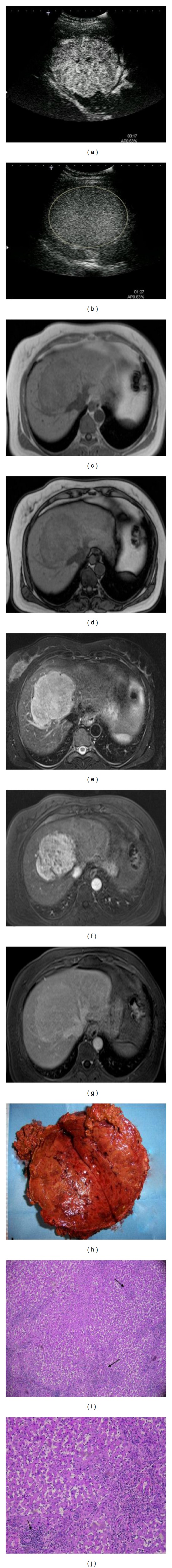
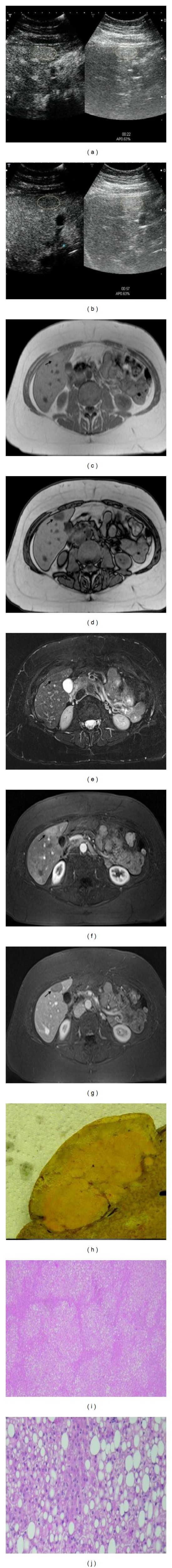
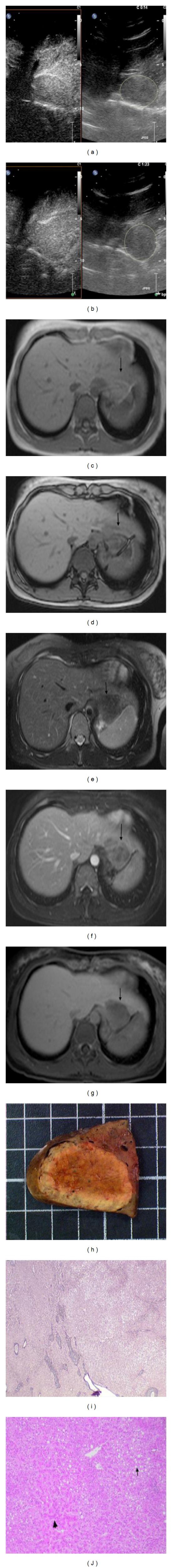
Purpose. To review the contrast-enhanced ultrasonographic (CEUS) and magnetic resonance (MR) imaging findings in 25 patients with 26 hepatocellular adenomas (HCAs) and to compare imaging features with histopathologic results from resected specimen considering the new immunophenotypical classification. Material and Methods. Two abdominal radiologists reviewed retrospectively CEUS cineloops and MR images in 26 HCA. All pathological specimens were reviewed and classified into four subgroups (steatotic or HNF 1α mutated, inflammatory, atypical or β-catenin mutated, and unspecified). Inflammatory infiltrates were scored, steatosis, and telangiectasia semiquantitatively evaluated. Results. CEUS and MRI features are well correlated: among the 16 inflammatory HCA, 7/16 presented typical imaging features: hypersignal T2, strong arterial enhancement with a centripetal filling, persistent on delayed phase. 6 HCA were classified as steatotic with typical imaging features: a drop out signal, slight arterial enhancement, vanishing on late phase. Four HCA were classified as atypical with an HCC developed in one. Five lesions displayed important steatosis (>50%) without belonging to the HNF1α group. Conclusion. In half cases, inflammatory HCA have specific imaging features well correlated with the amount of telangiectasia and inflammatory infiltrates. An HCA with important amount of steatosis noticed on chemical shift images does not always belong to the HNF1α group.

 求助内容:
求助内容: 应助结果提醒方式:


