P Lemkens, Gem Boari, Ge Fazzi, Gmj Janssen, Je Murphy-Ullrich, Pmh Schiffers, Jgr De Mey
{"title":"血栓反应蛋白-1在幼年正常血压大鼠和自发性高血压大鼠肠系膜动脉早期血流相关重构中的作用","authors":"P Lemkens, Gem Boari, Ge Fazzi, Gmj Janssen, Je Murphy-Ullrich, Pmh Schiffers, Jgr De Mey","doi":"10.2174/1874192401206010050","DOIUrl":null,"url":null,"abstract":"<p><p>We tested the hypotheses that TSP-1 participates in the initiation of remodeling of small muscular arteries in response to altered blood flow and that the N-terminal domain of TSP-1 (hepI) can reverse the pathological inward remodeling of resistance arteries from SHR.We measured (1) changes in gene/protein expression in MA of 6 week old WKY and SHR exposed to either increased (+ 100 %) or reduced blood flow (- 90 %) for 24-40 hours and (2) structural changes in MA of 12 week old SHR exposed for 3 days to hepI in organ culture.In both HF and LF of WKY, mRNA expression of eNOS, sGCα1 and PKG1β were significantly reduced (p < 0.05), whereas mRNA of TSP1 was markedly increased (p < 0.05). In MA of young SHR, similar results were obtained except that eNOS mRNA was not reduced in LF. Expression of TSP1 protein was significantly increased in LF of young WKY and SHR (p < 0.05). Exposure of MA of 12 week old SHR to hepI (1 µmol/L) resulted in a rapid lumen diameter increase (+ 12 ± 2% after 3 days) without alteration in vascular reactivity, distensibility, media surface area or cell number.These are the first observations of reduced gene expression of eNOS/sGC/PKG and increased expression of TSP1 at the initiation of arterial remodeling in young WKY and SHR, irrespective of its outward or inward outcome. Furthermore, a fragment of TSP-1 rapidly and directly reversed pathological inward arterial remodeling of SHR in vitro.</p>","PeriodicalId":504447,"journal":{"name":"The Open Cardiovascular Medicine Journal","volume":"6 ","pages":"50-9"},"PeriodicalIF":0.0000,"publicationDate":"2012-01-01","publicationTypes":"Journal Article","fieldsOfStudy":null,"isOpenAccess":false,"openAccessPdf":"https://ftp.ncbi.nlm.nih.gov/pub/pmc/oa_pdf/30/94/TOCMJ-6-50.PMC3367304.pdf","citationCount":"3","resultStr":"{\"title\":\"Thrombospondin-1 in early flow-related remodeling of mesenteric arteries from young normotensive and spontaneously hypertensive rats.\",\"authors\":\"P Lemkens, Gem Boari, Ge Fazzi, Gmj Janssen, Je Murphy-Ullrich, Pmh Schiffers, Jgr De Mey\",\"doi\":\"10.2174/1874192401206010050\",\"DOIUrl\":null,\"url\":null,\"abstract\":\"<p><p>We tested the hypotheses that TSP-1 participates in the initiation of remodeling of small muscular arteries in response to altered blood flow and that the N-terminal domain of TSP-1 (hepI) can reverse the pathological inward remodeling of resistance arteries from SHR.We measured (1) changes in gene/protein expression in MA of 6 week old WKY and SHR exposed to either increased (+ 100 %) or reduced blood flow (- 90 %) for 24-40 hours and (2) structural changes in MA of 12 week old SHR exposed for 3 days to hepI in organ culture.In both HF and LF of WKY, mRNA expression of eNOS, sGCα1 and PKG1β were significantly reduced (p < 0.05), whereas mRNA of TSP1 was markedly increased (p < 0.05). In MA of young SHR, similar results were obtained except that eNOS mRNA was not reduced in LF. Expression of TSP1 protein was significantly increased in LF of young WKY and SHR (p < 0.05). Exposure of MA of 12 week old SHR to hepI (1 µmol/L) resulted in a rapid lumen diameter increase (+ 12 ± 2% after 3 days) without alteration in vascular reactivity, distensibility, media surface area or cell number.These are the first observations of reduced gene expression of eNOS/sGC/PKG and increased expression of TSP1 at the initiation of arterial remodeling in young WKY and SHR, irrespective of its outward or inward outcome. Furthermore, a fragment of TSP-1 rapidly and directly reversed pathological inward arterial remodeling of SHR in vitro.</p>\",\"PeriodicalId\":504447,\"journal\":{\"name\":\"The Open Cardiovascular Medicine Journal\",\"volume\":\"6 \",\"pages\":\"50-9\"},\"PeriodicalIF\":0.0000,\"publicationDate\":\"2012-01-01\",\"publicationTypes\":\"Journal Article\",\"fieldsOfStudy\":null,\"isOpenAccess\":false,\"openAccessPdf\":\"https://ftp.ncbi.nlm.nih.gov/pub/pmc/oa_pdf/30/94/TOCMJ-6-50.PMC3367304.pdf\",\"citationCount\":\"3\",\"resultStr\":null,\"platform\":\"Semanticscholar\",\"paperid\":null,\"PeriodicalName\":\"The Open Cardiovascular Medicine Journal\",\"FirstCategoryId\":\"1085\",\"ListUrlMain\":\"https://doi.org/10.2174/1874192401206010050\",\"RegionNum\":0,\"RegionCategory\":null,\"ArticlePicture\":[],\"TitleCN\":null,\"AbstractTextCN\":null,\"PMCID\":null,\"EPubDate\":\"2012/5/18 0:00:00\",\"PubModel\":\"Epub\",\"JCR\":\"\",\"JCRName\":\"\",\"Score\":null,\"Total\":0}","platform":"Semanticscholar","paperid":null,"PeriodicalName":"The Open Cardiovascular Medicine Journal","FirstCategoryId":"1085","ListUrlMain":"https://doi.org/10.2174/1874192401206010050","RegionNum":0,"RegionCategory":null,"ArticlePicture":[],"TitleCN":null,"AbstractTextCN":null,"PMCID":null,"EPubDate":"2012/5/18 0:00:00","PubModel":"Epub","JCR":"","JCRName":"","Score":null,"Total":0}
Thrombospondin-1 in early flow-related remodeling of mesenteric arteries from young normotensive and spontaneously hypertensive rats.
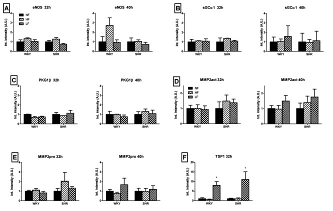
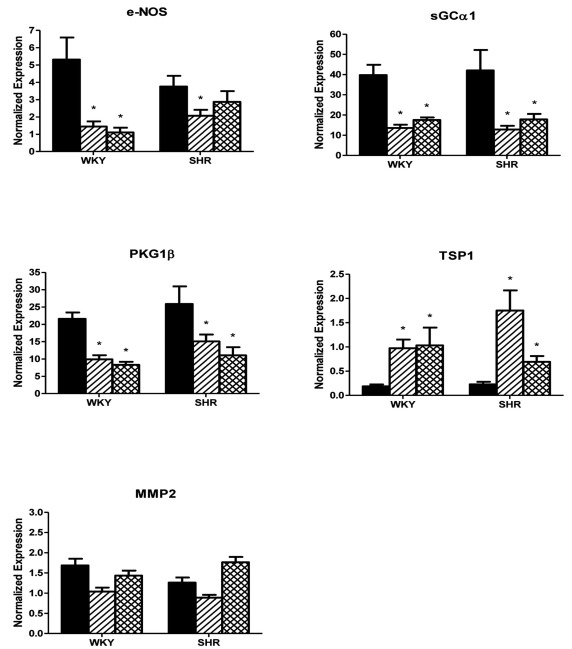
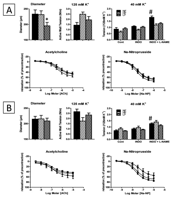
We tested the hypotheses that TSP-1 participates in the initiation of remodeling of small muscular arteries in response to altered blood flow and that the N-terminal domain of TSP-1 (hepI) can reverse the pathological inward remodeling of resistance arteries from SHR.We measured (1) changes in gene/protein expression in MA of 6 week old WKY and SHR exposed to either increased (+ 100 %) or reduced blood flow (- 90 %) for 24-40 hours and (2) structural changes in MA of 12 week old SHR exposed for 3 days to hepI in organ culture.In both HF and LF of WKY, mRNA expression of eNOS, sGCα1 and PKG1β were significantly reduced (p < 0.05), whereas mRNA of TSP1 was markedly increased (p < 0.05). In MA of young SHR, similar results were obtained except that eNOS mRNA was not reduced in LF. Expression of TSP1 protein was significantly increased in LF of young WKY and SHR (p < 0.05). Exposure of MA of 12 week old SHR to hepI (1 µmol/L) resulted in a rapid lumen diameter increase (+ 12 ± 2% after 3 days) without alteration in vascular reactivity, distensibility, media surface area or cell number.These are the first observations of reduced gene expression of eNOS/sGC/PKG and increased expression of TSP1 at the initiation of arterial remodeling in young WKY and SHR, irrespective of its outward or inward outcome. Furthermore, a fragment of TSP-1 rapidly and directly reversed pathological inward arterial remodeling of SHR in vitro.

 求助内容:
求助内容: 应助结果提醒方式:


