Kyu Sik Jung, Kyeong Hye Park, Young Eun Chon, Sa Ra Lee, Young Nyun Park, Do Yun Lee, Jin Sil Seong, Jun Yong Park
{"title":"孤立性转移性肝细胞癌发生于骨盆骨1例。","authors":"Kyu Sik Jung, Kyeong Hye Park, Young Eun Chon, Sa Ra Lee, Young Nyun Park, Do Yun Lee, Jin Sil Seong, Jun Yong Park","doi":"10.3350/kjhep.2012.18.1.89","DOIUrl":null,"url":null,"abstract":"<p><p>Reports of metastatic hepatocellular carcinoma (HCC) without a primary liver tumor are rare. Here we present a case of isolated HCC that had metastasized to the pelvic bone without a primary focus. A 73-year-old man presented with severe back and right-leg pain. Radiological examinations, including computed tomography (CT) and magnetic resonance imaging (MRI), revealed a huge mass on the pelvic bone (13×10 cm). He underwent an incisional biopsy, and the results of the subsequent histological examination were consistent with metastatic hepatocellular carcinoma. The tumor cells were positive for cytokeratin (AE1/AE3), hepatocyte paraffin 1, and glypican-3, and negative for CD56, chromogranin A, and synaptophysin on immunohistochemical staining. Examination of the liver by CT, MRI, positron-emission tomography scan, and angiography produced no evidence of a primary tumor. Radiotherapy and transarterial chemoembolization were performed on the pelvic bone, followed by systemic chemotherapy. These combination treatments resulted in tumor regression with necrotic changes. However, multiple lung metastases developed 1 year after the treatment, and the patient was treated with additional systemic chemotherapy.</p>","PeriodicalId":87153,"journal":{"name":"The Korean journal of hepatology","volume":"18 1","pages":"89-93"},"PeriodicalIF":0.0000,"publicationDate":"2012-03-01","publicationTypes":"Journal Article","fieldsOfStudy":null,"isOpenAccess":false,"openAccessPdf":"https://ftp.ncbi.nlm.nih.gov/pub/pmc/oa_pdf/68/26/kjhep-18-89.PMC3326999.pdf","citationCount":"8","resultStr":"{\"title\":\"A case of isolated metastatic hepatocellular carcinoma arising from the pelvic bone.\",\"authors\":\"Kyu Sik Jung, Kyeong Hye Park, Young Eun Chon, Sa Ra Lee, Young Nyun Park, Do Yun Lee, Jin Sil Seong, Jun Yong Park\",\"doi\":\"10.3350/kjhep.2012.18.1.89\",\"DOIUrl\":null,\"url\":null,\"abstract\":\"<p><p>Reports of metastatic hepatocellular carcinoma (HCC) without a primary liver tumor are rare. Here we present a case of isolated HCC that had metastasized to the pelvic bone without a primary focus. A 73-year-old man presented with severe back and right-leg pain. Radiological examinations, including computed tomography (CT) and magnetic resonance imaging (MRI), revealed a huge mass on the pelvic bone (13×10 cm). He underwent an incisional biopsy, and the results of the subsequent histological examination were consistent with metastatic hepatocellular carcinoma. The tumor cells were positive for cytokeratin (AE1/AE3), hepatocyte paraffin 1, and glypican-3, and negative for CD56, chromogranin A, and synaptophysin on immunohistochemical staining. Examination of the liver by CT, MRI, positron-emission tomography scan, and angiography produced no evidence of a primary tumor. Radiotherapy and transarterial chemoembolization were performed on the pelvic bone, followed by systemic chemotherapy. These combination treatments resulted in tumor regression with necrotic changes. However, multiple lung metastases developed 1 year after the treatment, and the patient was treated with additional systemic chemotherapy.</p>\",\"PeriodicalId\":87153,\"journal\":{\"name\":\"The Korean journal of hepatology\",\"volume\":\"18 1\",\"pages\":\"89-93\"},\"PeriodicalIF\":0.0000,\"publicationDate\":\"2012-03-01\",\"publicationTypes\":\"Journal Article\",\"fieldsOfStudy\":null,\"isOpenAccess\":false,\"openAccessPdf\":\"https://ftp.ncbi.nlm.nih.gov/pub/pmc/oa_pdf/68/26/kjhep-18-89.PMC3326999.pdf\",\"citationCount\":\"8\",\"resultStr\":null,\"platform\":\"Semanticscholar\",\"paperid\":null,\"PeriodicalName\":\"The Korean journal of hepatology\",\"FirstCategoryId\":\"1085\",\"ListUrlMain\":\"https://doi.org/10.3350/kjhep.2012.18.1.89\",\"RegionNum\":0,\"RegionCategory\":null,\"ArticlePicture\":[],\"TitleCN\":null,\"AbstractTextCN\":null,\"PMCID\":null,\"EPubDate\":\"2012/3/22 0:00:00\",\"PubModel\":\"Epub\",\"JCR\":\"\",\"JCRName\":\"\",\"Score\":null,\"Total\":0}","platform":"Semanticscholar","paperid":null,"PeriodicalName":"The Korean journal of hepatology","FirstCategoryId":"1085","ListUrlMain":"https://doi.org/10.3350/kjhep.2012.18.1.89","RegionNum":0,"RegionCategory":null,"ArticlePicture":[],"TitleCN":null,"AbstractTextCN":null,"PMCID":null,"EPubDate":"2012/3/22 0:00:00","PubModel":"Epub","JCR":"","JCRName":"","Score":null,"Total":0}
A case of isolated metastatic hepatocellular carcinoma arising from the pelvic bone.
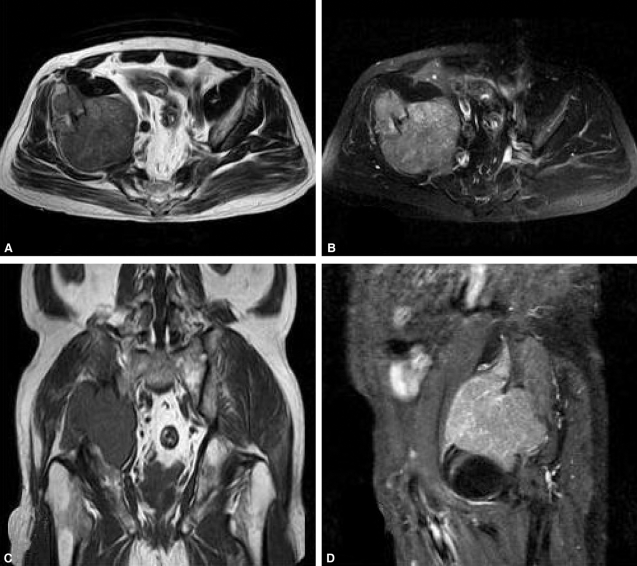
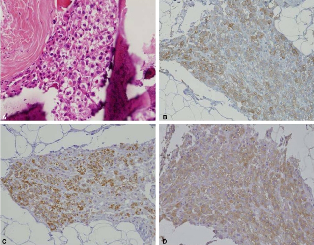
Reports of metastatic hepatocellular carcinoma (HCC) without a primary liver tumor are rare. Here we present a case of isolated HCC that had metastasized to the pelvic bone without a primary focus. A 73-year-old man presented with severe back and right-leg pain. Radiological examinations, including computed tomography (CT) and magnetic resonance imaging (MRI), revealed a huge mass on the pelvic bone (13×10 cm). He underwent an incisional biopsy, and the results of the subsequent histological examination were consistent with metastatic hepatocellular carcinoma. The tumor cells were positive for cytokeratin (AE1/AE3), hepatocyte paraffin 1, and glypican-3, and negative for CD56, chromogranin A, and synaptophysin on immunohistochemical staining. Examination of the liver by CT, MRI, positron-emission tomography scan, and angiography produced no evidence of a primary tumor. Radiotherapy and transarterial chemoembolization were performed on the pelvic bone, followed by systemic chemotherapy. These combination treatments resulted in tumor regression with necrotic changes. However, multiple lung metastases developed 1 year after the treatment, and the patient was treated with additional systemic chemotherapy.

 求助内容:
求助内容: 应助结果提醒方式:


