Julie A Chouinard, Jacques A Rousseau, Jean-François Beaudoin, Patrick Vermette, Roger Lecomte
{"title":"人内皮细胞和成纤维细胞单层正电子发射断层扫描检测:预处理和细胞密度对18FDG摄取的影响。","authors":"Julie A Chouinard, Jacques A Rousseau, Jean-François Beaudoin, Patrick Vermette, Roger Lecomte","doi":"10.1186/2045-824X-4-5","DOIUrl":null,"url":null,"abstract":"<p><strong>Background: </strong>The non-destructive assessment and characterization of tridimensional (3D) cell and tissue constructs in bioreactors represents a challenge in tissue engineering. Medical imaging modalities, which can provide information on the structure and function of internal organs and tissues in living organisms, have the potential of allowing repetitive monitoring of these 3D cultures in vitro. Positron emission tomography (PET) is the most sensitive non-invasive imaging modality, capable of measuring picomolar amounts of radiolabeled molecules. However, since PET imaging protocols have been designed almost exclusively for in vivo investigations, suitable methods must be devised to enable imaging of cells or tissue substitutes. As a prior step to imaging 3D cultures, cell radiotracer uptake conditions must first be optimized.</p><p><strong>Methods: </strong>In this study, human umbilical vein endothelial cells (HUVEC) and human fibroblasts were cultured at different densities and PET was used to non-destructively monitor their glycolytic activity by measuring 18F-fluorodeoxyglucose (18FDG) uptake. Various cell preconditioning protocols were investigated by adjusting the following parameters to optimize 18FDG uptake: glucose starvation, insulin stimulation, glucose concentration, 18FDG incubation time, cell density and radiotracer efflux prevention.</p><p><strong>Results: </strong>The conditions yielding optimal 18FDG uptake, and hence best detection sensitivity by PET, were as follows: 2-hour cell preconditioning by glucose deprivation with 1-hour insulin stimulation, followed by 1-hour 18FDG incubation and 15-minute stabilization in standard culture medium, prior to rinsing and PET scanning.</p><p><strong>Conclusions: </strong>A step-wise dependence of 18FDG uptake on glucose concentration was found, but a linear correlation between PET signal and cell density was observed. Detection thresholds of 36 ± 7 and 21 ± 4 cells were estimated for endothelial cells and fibroblasts, respectively.</p>","PeriodicalId":23948,"journal":{"name":"Vascular Cell","volume":"4 1","pages":"5"},"PeriodicalIF":0.0000,"publicationDate":"2012-03-20","publicationTypes":"Journal Article","fieldsOfStudy":null,"isOpenAccess":false,"openAccessPdf":"https://sci-hub-pdf.com/10.1186/2045-824X-4-5","citationCount":"13","resultStr":"{\"title\":\"Positron emission tomography detection of human endothelial cell and fibroblast monolayers: effect of pretreament and cell density on 18FDG uptake.\",\"authors\":\"Julie A Chouinard, Jacques A Rousseau, Jean-François Beaudoin, Patrick Vermette, Roger Lecomte\",\"doi\":\"10.1186/2045-824X-4-5\",\"DOIUrl\":null,\"url\":null,\"abstract\":\"<p><strong>Background: </strong>The non-destructive assessment and characterization of tridimensional (3D) cell and tissue constructs in bioreactors represents a challenge in tissue engineering. Medical imaging modalities, which can provide information on the structure and function of internal organs and tissues in living organisms, have the potential of allowing repetitive monitoring of these 3D cultures in vitro. Positron emission tomography (PET) is the most sensitive non-invasive imaging modality, capable of measuring picomolar amounts of radiolabeled molecules. However, since PET imaging protocols have been designed almost exclusively for in vivo investigations, suitable methods must be devised to enable imaging of cells or tissue substitutes. As a prior step to imaging 3D cultures, cell radiotracer uptake conditions must first be optimized.</p><p><strong>Methods: </strong>In this study, human umbilical vein endothelial cells (HUVEC) and human fibroblasts were cultured at different densities and PET was used to non-destructively monitor their glycolytic activity by measuring 18F-fluorodeoxyglucose (18FDG) uptake. Various cell preconditioning protocols were investigated by adjusting the following parameters to optimize 18FDG uptake: glucose starvation, insulin stimulation, glucose concentration, 18FDG incubation time, cell density and radiotracer efflux prevention.</p><p><strong>Results: </strong>The conditions yielding optimal 18FDG uptake, and hence best detection sensitivity by PET, were as follows: 2-hour cell preconditioning by glucose deprivation with 1-hour insulin stimulation, followed by 1-hour 18FDG incubation and 15-minute stabilization in standard culture medium, prior to rinsing and PET scanning.</p><p><strong>Conclusions: </strong>A step-wise dependence of 18FDG uptake on glucose concentration was found, but a linear correlation between PET signal and cell density was observed. Detection thresholds of 36 ± 7 and 21 ± 4 cells were estimated for endothelial cells and fibroblasts, respectively.</p>\",\"PeriodicalId\":23948,\"journal\":{\"name\":\"Vascular Cell\",\"volume\":\"4 1\",\"pages\":\"5\"},\"PeriodicalIF\":0.0000,\"publicationDate\":\"2012-03-20\",\"publicationTypes\":\"Journal Article\",\"fieldsOfStudy\":null,\"isOpenAccess\":false,\"openAccessPdf\":\"https://sci-hub-pdf.com/10.1186/2045-824X-4-5\",\"citationCount\":\"13\",\"resultStr\":null,\"platform\":\"Semanticscholar\",\"paperid\":null,\"PeriodicalName\":\"Vascular Cell\",\"FirstCategoryId\":\"1085\",\"ListUrlMain\":\"https://doi.org/10.1186/2045-824X-4-5\",\"RegionNum\":0,\"RegionCategory\":null,\"ArticlePicture\":[],\"TitleCN\":null,\"AbstractTextCN\":null,\"PMCID\":null,\"EPubDate\":\"\",\"PubModel\":\"\",\"JCR\":\"Q4\",\"JCRName\":\"Neuroscience\",\"Score\":null,\"Total\":0}","platform":"Semanticscholar","paperid":null,"PeriodicalName":"Vascular Cell","FirstCategoryId":"1085","ListUrlMain":"https://doi.org/10.1186/2045-824X-4-5","RegionNum":0,"RegionCategory":null,"ArticlePicture":[],"TitleCN":null,"AbstractTextCN":null,"PMCID":null,"EPubDate":"","PubModel":"","JCR":"Q4","JCRName":"Neuroscience","Score":null,"Total":0}
Positron emission tomography detection of human endothelial cell and fibroblast monolayers: effect of pretreament and cell density on 18FDG uptake.
Background: The non-destructive assessment and characterization of tridimensional (3D) cell and tissue constructs in bioreactors represents a challenge in tissue engineering. Medical imaging modalities, which can provide information on the structure and function of internal organs and tissues in living organisms, have the potential of allowing repetitive monitoring of these 3D cultures in vitro. Positron emission tomography (PET) is the most sensitive non-invasive imaging modality, capable of measuring picomolar amounts of radiolabeled molecules. However, since PET imaging protocols have been designed almost exclusively for in vivo investigations, suitable methods must be devised to enable imaging of cells or tissue substitutes. As a prior step to imaging 3D cultures, cell radiotracer uptake conditions must first be optimized.
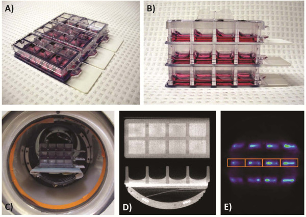
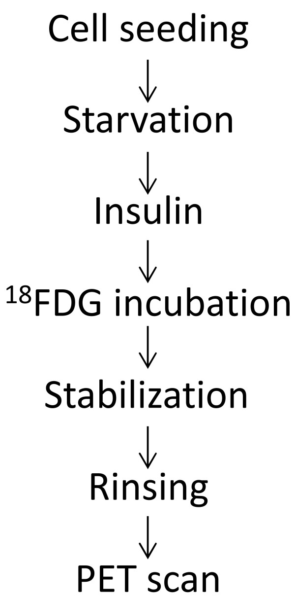
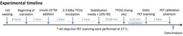
Methods: In this study, human umbilical vein endothelial cells (HUVEC) and human fibroblasts were cultured at different densities and PET was used to non-destructively monitor their glycolytic activity by measuring 18F-fluorodeoxyglucose (18FDG) uptake. Various cell preconditioning protocols were investigated by adjusting the following parameters to optimize 18FDG uptake: glucose starvation, insulin stimulation, glucose concentration, 18FDG incubation time, cell density and radiotracer efflux prevention.
Results: The conditions yielding optimal 18FDG uptake, and hence best detection sensitivity by PET, were as follows: 2-hour cell preconditioning by glucose deprivation with 1-hour insulin stimulation, followed by 1-hour 18FDG incubation and 15-minute stabilization in standard culture medium, prior to rinsing and PET scanning.
Conclusions: A step-wise dependence of 18FDG uptake on glucose concentration was found, but a linear correlation between PET signal and cell density was observed. Detection thresholds of 36 ± 7 and 21 ± 4 cells were estimated for endothelial cells and fibroblasts, respectively.

 求助内容:
求助内容: 应助结果提醒方式:


