{"title":"基于数学形态学的医学图像形态学特征增强方法。","authors":"Yoshitaka Kimori","doi":"10.1186/2043-9113-1-33","DOIUrl":null,"url":null,"abstract":"<p><strong>Background: </strong>Medical image processing is essential in many fields of medical research and clinical practice because it greatly facilitates early and accurate detection and diagnosis of diseases. In particular, contrast enhancement is important for optimal image quality and visibility. This paper proposes a new image processing method for enhancing morphological features of masses and other abnormalities in medical images.</p><p><strong>Method: </strong>The proposed method involves two steps: (1) selective extraction of target features by mathematical morphology and (2) enhancement of the extracted features by two contrast modification techniques.</p><p><strong>Results: </strong>The goal of the proposed method is to enable enhancement of fine morphological features of a lesion region with high suppression of surrounding tissues. The effectiveness of the method was evaluated in quantitative terms of the contrast improvement ratio. The results clearly show that the method outperforms five conventional contrast enhancement methods. The effectiveness and usefulness of the proposed method were further demonstrated by application to three types of medical images: a mammographic image, a chest radiographic image, and a retinal image.</p><p><strong>Conclusion: </strong>The proposed method enables specific extraction and enhancement of mass lesions, which is essential for clinical diagnosis based on medical image analysis. Thus, the method can be expected to achieve automatic recognition of lesion location and quantitative analysis of legion morphology.</p>","PeriodicalId":73663,"journal":{"name":"Journal of clinical bioinformatics","volume":"1 ","pages":"33"},"PeriodicalIF":0.0000,"publicationDate":"2011-12-16","publicationTypes":"Journal Article","fieldsOfStudy":null,"isOpenAccess":false,"openAccessPdf":"https://sci-hub-pdf.com/10.1186/2043-9113-1-33","citationCount":"68","resultStr":"{\"title\":\"Mathematical morphology-based approach to the enhancement of morphological features in medical images.\",\"authors\":\"Yoshitaka Kimori\",\"doi\":\"10.1186/2043-9113-1-33\",\"DOIUrl\":null,\"url\":null,\"abstract\":\"<p><strong>Background: </strong>Medical image processing is essential in many fields of medical research and clinical practice because it greatly facilitates early and accurate detection and diagnosis of diseases. In particular, contrast enhancement is important for optimal image quality and visibility. This paper proposes a new image processing method for enhancing morphological features of masses and other abnormalities in medical images.</p><p><strong>Method: </strong>The proposed method involves two steps: (1) selective extraction of target features by mathematical morphology and (2) enhancement of the extracted features by two contrast modification techniques.</p><p><strong>Results: </strong>The goal of the proposed method is to enable enhancement of fine morphological features of a lesion region with high suppression of surrounding tissues. The effectiveness of the method was evaluated in quantitative terms of the contrast improvement ratio. The results clearly show that the method outperforms five conventional contrast enhancement methods. The effectiveness and usefulness of the proposed method were further demonstrated by application to three types of medical images: a mammographic image, a chest radiographic image, and a retinal image.</p><p><strong>Conclusion: </strong>The proposed method enables specific extraction and enhancement of mass lesions, which is essential for clinical diagnosis based on medical image analysis. Thus, the method can be expected to achieve automatic recognition of lesion location and quantitative analysis of legion morphology.</p>\",\"PeriodicalId\":73663,\"journal\":{\"name\":\"Journal of clinical bioinformatics\",\"volume\":\"1 \",\"pages\":\"33\"},\"PeriodicalIF\":0.0000,\"publicationDate\":\"2011-12-16\",\"publicationTypes\":\"Journal Article\",\"fieldsOfStudy\":null,\"isOpenAccess\":false,\"openAccessPdf\":\"https://sci-hub-pdf.com/10.1186/2043-9113-1-33\",\"citationCount\":\"68\",\"resultStr\":null,\"platform\":\"Semanticscholar\",\"paperid\":null,\"PeriodicalName\":\"Journal of clinical bioinformatics\",\"FirstCategoryId\":\"1085\",\"ListUrlMain\":\"https://doi.org/10.1186/2043-9113-1-33\",\"RegionNum\":0,\"RegionCategory\":null,\"ArticlePicture\":[],\"TitleCN\":null,\"AbstractTextCN\":null,\"PMCID\":null,\"EPubDate\":\"\",\"PubModel\":\"\",\"JCR\":\"\",\"JCRName\":\"\",\"Score\":null,\"Total\":0}","platform":"Semanticscholar","paperid":null,"PeriodicalName":"Journal of clinical bioinformatics","FirstCategoryId":"1085","ListUrlMain":"https://doi.org/10.1186/2043-9113-1-33","RegionNum":0,"RegionCategory":null,"ArticlePicture":[],"TitleCN":null,"AbstractTextCN":null,"PMCID":null,"EPubDate":"","PubModel":"","JCR":"","JCRName":"","Score":null,"Total":0}
Mathematical morphology-based approach to the enhancement of morphological features in medical images.
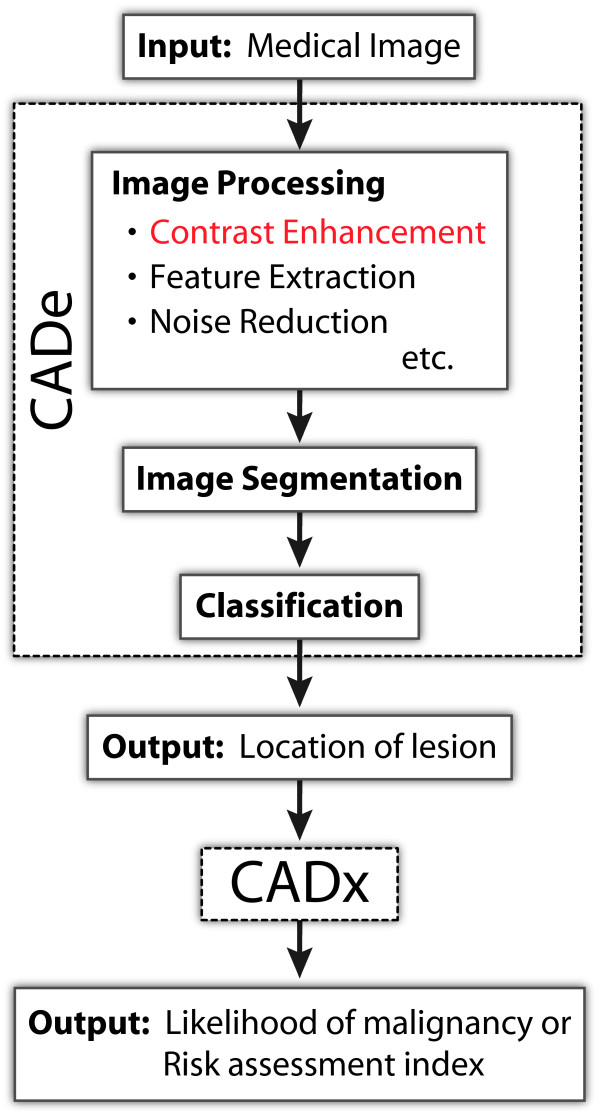
Background: Medical image processing is essential in many fields of medical research and clinical practice because it greatly facilitates early and accurate detection and diagnosis of diseases. In particular, contrast enhancement is important for optimal image quality and visibility. This paper proposes a new image processing method for enhancing morphological features of masses and other abnormalities in medical images.
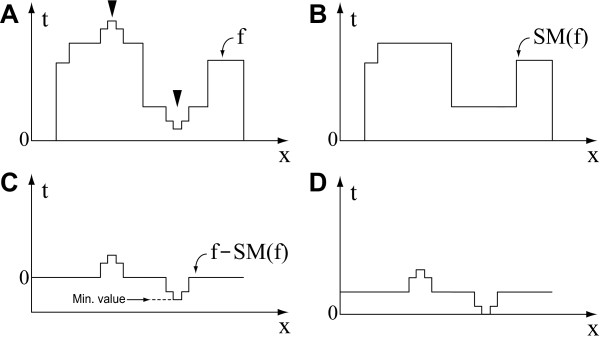
Method: The proposed method involves two steps: (1) selective extraction of target features by mathematical morphology and (2) enhancement of the extracted features by two contrast modification techniques.
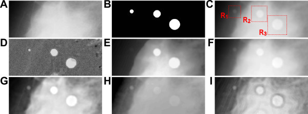
Results: The goal of the proposed method is to enable enhancement of fine morphological features of a lesion region with high suppression of surrounding tissues. The effectiveness of the method was evaluated in quantitative terms of the contrast improvement ratio. The results clearly show that the method outperforms five conventional contrast enhancement methods. The effectiveness and usefulness of the proposed method were further demonstrated by application to three types of medical images: a mammographic image, a chest radiographic image, and a retinal image.
Conclusion: The proposed method enables specific extraction and enhancement of mass lesions, which is essential for clinical diagnosis based on medical image analysis. Thus, the method can be expected to achieve automatic recognition of lesion location and quantitative analysis of legion morphology.

 求助内容:
求助内容: 应助结果提醒方式:


