Julie C Williams, Rebecca D Lee, Claire M Doerschuk, Nigel Mackman
{"title":"pa -2缺乏对小鼠气管内LPS处理后KC表达的影响。","authors":"Julie C Williams, Rebecca D Lee, Claire M Doerschuk, Nigel Mackman","doi":"10.1155/2011/415195","DOIUrl":null,"url":null,"abstract":"<p><p>Protease activated receptors (PAR) have been shown to play a role in inflammation. PAR-2 is expressed by numerous cells in the lung and has either proinflammatory, anti-inflammatory, or no effect depending on the model. Here, we examined the role of PAR-2 in a model of LPS-induced lung inflammation. We found that PAR-2-deficient mice had significantly less KC expression in bronchial lavage fluid compared with wild-type mice but there was no difference in MIP-2 or TNF-α expression. We also found that isolated alveolar and resident peritoneal macrophages lacking PAR-2 showed a similar deficit in KC after LPS stimulation without differences in MIP-2 or TNF-α. Infiltration of neutrophils and macrophages into the lung following LPS administration was not affected by an absence of PAR-2. Our results support the notion that PAR-2 plays a role in LPS activation of TLR4 signaling in macrophages.</p>","PeriodicalId":89176,"journal":{"name":"Journal of signal transduction","volume":"2011 ","pages":"415195"},"PeriodicalIF":0.0000,"publicationDate":"2011-01-01","publicationTypes":"Journal Article","fieldsOfStudy":null,"isOpenAccess":false,"openAccessPdf":"https://sci-hub-pdf.com/10.1155/2011/415195","citationCount":"12","resultStr":"{\"title\":\"Effect of PAR-2 Deficiency in Mice on KC Expression after Intratracheal LPS Administration.\",\"authors\":\"Julie C Williams, Rebecca D Lee, Claire M Doerschuk, Nigel Mackman\",\"doi\":\"10.1155/2011/415195\",\"DOIUrl\":null,\"url\":null,\"abstract\":\"<p><p>Protease activated receptors (PAR) have been shown to play a role in inflammation. PAR-2 is expressed by numerous cells in the lung and has either proinflammatory, anti-inflammatory, or no effect depending on the model. Here, we examined the role of PAR-2 in a model of LPS-induced lung inflammation. We found that PAR-2-deficient mice had significantly less KC expression in bronchial lavage fluid compared with wild-type mice but there was no difference in MIP-2 or TNF-α expression. We also found that isolated alveolar and resident peritoneal macrophages lacking PAR-2 showed a similar deficit in KC after LPS stimulation without differences in MIP-2 or TNF-α. Infiltration of neutrophils and macrophages into the lung following LPS administration was not affected by an absence of PAR-2. Our results support the notion that PAR-2 plays a role in LPS activation of TLR4 signaling in macrophages.</p>\",\"PeriodicalId\":89176,\"journal\":{\"name\":\"Journal of signal transduction\",\"volume\":\"2011 \",\"pages\":\"415195\"},\"PeriodicalIF\":0.0000,\"publicationDate\":\"2011-01-01\",\"publicationTypes\":\"Journal Article\",\"fieldsOfStudy\":null,\"isOpenAccess\":false,\"openAccessPdf\":\"https://sci-hub-pdf.com/10.1155/2011/415195\",\"citationCount\":\"12\",\"resultStr\":null,\"platform\":\"Semanticscholar\",\"paperid\":null,\"PeriodicalName\":\"Journal of signal transduction\",\"FirstCategoryId\":\"1085\",\"ListUrlMain\":\"https://doi.org/10.1155/2011/415195\",\"RegionNum\":0,\"RegionCategory\":null,\"ArticlePicture\":[],\"TitleCN\":null,\"AbstractTextCN\":null,\"PMCID\":null,\"EPubDate\":\"2011/12/7 0:00:00\",\"PubModel\":\"Epub\",\"JCR\":\"\",\"JCRName\":\"\",\"Score\":null,\"Total\":0}","platform":"Semanticscholar","paperid":null,"PeriodicalName":"Journal of signal transduction","FirstCategoryId":"1085","ListUrlMain":"https://doi.org/10.1155/2011/415195","RegionNum":0,"RegionCategory":null,"ArticlePicture":[],"TitleCN":null,"AbstractTextCN":null,"PMCID":null,"EPubDate":"2011/12/7 0:00:00","PubModel":"Epub","JCR":"","JCRName":"","Score":null,"Total":0}
Effect of PAR-2 Deficiency in Mice on KC Expression after Intratracheal LPS Administration.
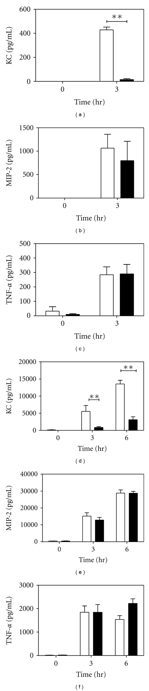
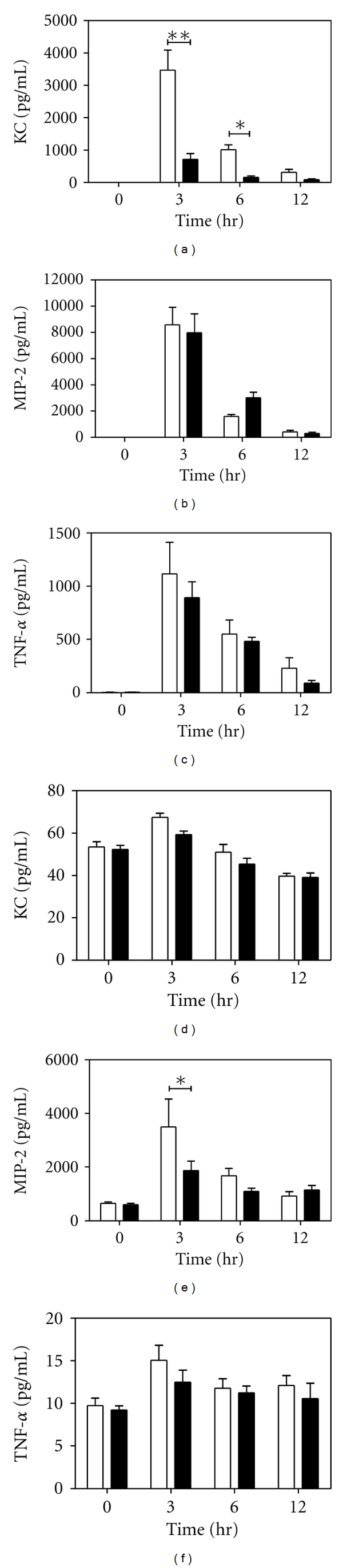
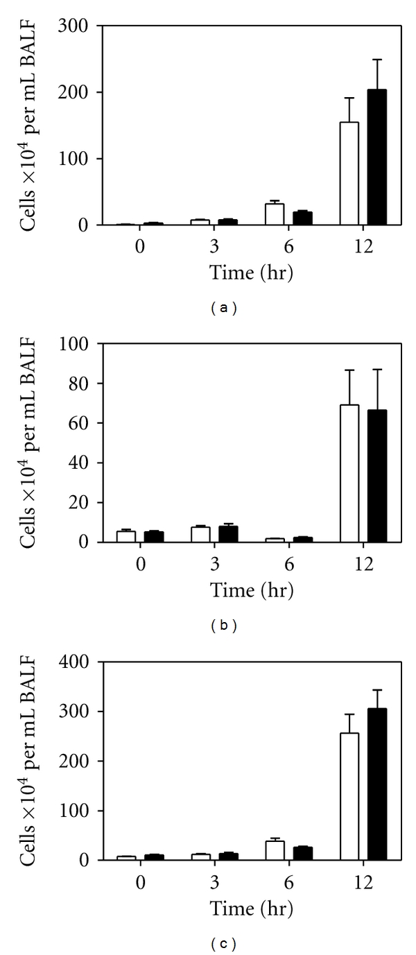
Protease activated receptors (PAR) have been shown to play a role in inflammation. PAR-2 is expressed by numerous cells in the lung and has either proinflammatory, anti-inflammatory, or no effect depending on the model. Here, we examined the role of PAR-2 in a model of LPS-induced lung inflammation. We found that PAR-2-deficient mice had significantly less KC expression in bronchial lavage fluid compared with wild-type mice but there was no difference in MIP-2 or TNF-α expression. We also found that isolated alveolar and resident peritoneal macrophages lacking PAR-2 showed a similar deficit in KC after LPS stimulation without differences in MIP-2 or TNF-α. Infiltration of neutrophils and macrophages into the lung following LPS administration was not affected by an absence of PAR-2. Our results support the notion that PAR-2 plays a role in LPS activation of TLR4 signaling in macrophages.

 求助内容:
求助内容: 应助结果提醒方式:


