Claudia Landi, Elena Bargagli, Barbara Magi, Antje Prasse, Joachim Muller-Quernheim, Luca Bini, Paola Rottoli
{"title":"肺朗格汉斯细胞组织细胞增多症支气管肺泡灌洗的蛋白质组学分析。","authors":"Claudia Landi, Elena Bargagli, Barbara Magi, Antje Prasse, Joachim Muller-Quernheim, Luca Bini, Paola Rottoli","doi":"10.1186/2043-9113-1-31","DOIUrl":null,"url":null,"abstract":"<p><strong>Background: </strong>Pulmonary Langerhans-cell histiocytosis (PLCH) is a rare interstitial lung disease characterized by clusters of Langerhans cells, organized in granulomas, in the walls of distal bronchioles. It is a diffuse lung disease related to tobacco smoking but otherwise of unknown etiopathogenesis.</p><p><strong>Methods: </strong>In this study we used a proteomic approach to analyze BAL protein composition of patients with PLCH and of healthy smoker and non-smoker controls to obtain insights into the pathogenetic mechanisms of the disease, to study the effect of cigarette smoking on susceptibility to PLCH and to identify potential new biomarkers.</p><p><strong>Results: </strong>Two-dimensional electrophoresis and image analysis revealed proteins that were differently expressed (quantitatively and qualitatively) in the three groups of subjects. The proteins were identified by mass spectrometry and have various functions (antioxidant, proinflammatory, antiprotease) and origins (plasma, locally produced, etc.). Many, such as protease inhibitors (human serpin B3) and antioxidant proteins (glutathione peroxidase and thioredoxin) are already linked to PLCH pathogenesis, whereas other proteins have never been associated with the disease. Interestingly, numerous proteolytic fragments of plasma proteins (including kininogen-1 N fragments and haptoglobin) were also identified and suggest increased proteolytic activity in this inflammatory lung disease. Differences in protein expression were found between the three groups and confirmed by Principal Component Analysis (PCA).</p><p><strong>Conclusion: </strong>Analysis of BAL proteomes of PLCH patients and of smoker and non-smoker controls also proved to be useful for researching the pathogenetic mechanisms and for identifying biomarkers of this rare diffuse lung disease.</p>","PeriodicalId":73663,"journal":{"name":"Journal of clinical bioinformatics","volume":"1 ","pages":"31"},"PeriodicalIF":0.0000,"publicationDate":"2011-11-10","publicationTypes":"Journal Article","fieldsOfStudy":null,"isOpenAccess":false,"openAccessPdf":"https://sci-hub-pdf.com/10.1186/2043-9113-1-31","citationCount":"22","resultStr":"{\"title\":\"Proteome analysis of bronchoalveolar lavage in pulmonary langerhans cell histiocytosis.\",\"authors\":\"Claudia Landi, Elena Bargagli, Barbara Magi, Antje Prasse, Joachim Muller-Quernheim, Luca Bini, Paola Rottoli\",\"doi\":\"10.1186/2043-9113-1-31\",\"DOIUrl\":null,\"url\":null,\"abstract\":\"<p><strong>Background: </strong>Pulmonary Langerhans-cell histiocytosis (PLCH) is a rare interstitial lung disease characterized by clusters of Langerhans cells, organized in granulomas, in the walls of distal bronchioles. It is a diffuse lung disease related to tobacco smoking but otherwise of unknown etiopathogenesis.</p><p><strong>Methods: </strong>In this study we used a proteomic approach to analyze BAL protein composition of patients with PLCH and of healthy smoker and non-smoker controls to obtain insights into the pathogenetic mechanisms of the disease, to study the effect of cigarette smoking on susceptibility to PLCH and to identify potential new biomarkers.</p><p><strong>Results: </strong>Two-dimensional electrophoresis and image analysis revealed proteins that were differently expressed (quantitatively and qualitatively) in the three groups of subjects. The proteins were identified by mass spectrometry and have various functions (antioxidant, proinflammatory, antiprotease) and origins (plasma, locally produced, etc.). Many, such as protease inhibitors (human serpin B3) and antioxidant proteins (glutathione peroxidase and thioredoxin) are already linked to PLCH pathogenesis, whereas other proteins have never been associated with the disease. Interestingly, numerous proteolytic fragments of plasma proteins (including kininogen-1 N fragments and haptoglobin) were also identified and suggest increased proteolytic activity in this inflammatory lung disease. Differences in protein expression were found between the three groups and confirmed by Principal Component Analysis (PCA).</p><p><strong>Conclusion: </strong>Analysis of BAL proteomes of PLCH patients and of smoker and non-smoker controls also proved to be useful for researching the pathogenetic mechanisms and for identifying biomarkers of this rare diffuse lung disease.</p>\",\"PeriodicalId\":73663,\"journal\":{\"name\":\"Journal of clinical bioinformatics\",\"volume\":\"1 \",\"pages\":\"31\"},\"PeriodicalIF\":0.0000,\"publicationDate\":\"2011-11-10\",\"publicationTypes\":\"Journal Article\",\"fieldsOfStudy\":null,\"isOpenAccess\":false,\"openAccessPdf\":\"https://sci-hub-pdf.com/10.1186/2043-9113-1-31\",\"citationCount\":\"22\",\"resultStr\":null,\"platform\":\"Semanticscholar\",\"paperid\":null,\"PeriodicalName\":\"Journal of clinical bioinformatics\",\"FirstCategoryId\":\"1085\",\"ListUrlMain\":\"https://doi.org/10.1186/2043-9113-1-31\",\"RegionNum\":0,\"RegionCategory\":null,\"ArticlePicture\":[],\"TitleCN\":null,\"AbstractTextCN\":null,\"PMCID\":null,\"EPubDate\":\"\",\"PubModel\":\"\",\"JCR\":\"\",\"JCRName\":\"\",\"Score\":null,\"Total\":0}","platform":"Semanticscholar","paperid":null,"PeriodicalName":"Journal of clinical bioinformatics","FirstCategoryId":"1085","ListUrlMain":"https://doi.org/10.1186/2043-9113-1-31","RegionNum":0,"RegionCategory":null,"ArticlePicture":[],"TitleCN":null,"AbstractTextCN":null,"PMCID":null,"EPubDate":"","PubModel":"","JCR":"","JCRName":"","Score":null,"Total":0}
Proteome analysis of bronchoalveolar lavage in pulmonary langerhans cell histiocytosis.
Background: Pulmonary Langerhans-cell histiocytosis (PLCH) is a rare interstitial lung disease characterized by clusters of Langerhans cells, organized in granulomas, in the walls of distal bronchioles. It is a diffuse lung disease related to tobacco smoking but otherwise of unknown etiopathogenesis.
Methods: In this study we used a proteomic approach to analyze BAL protein composition of patients with PLCH and of healthy smoker and non-smoker controls to obtain insights into the pathogenetic mechanisms of the disease, to study the effect of cigarette smoking on susceptibility to PLCH and to identify potential new biomarkers.
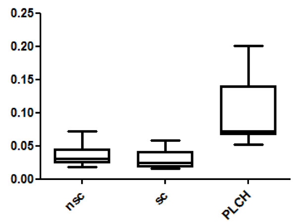
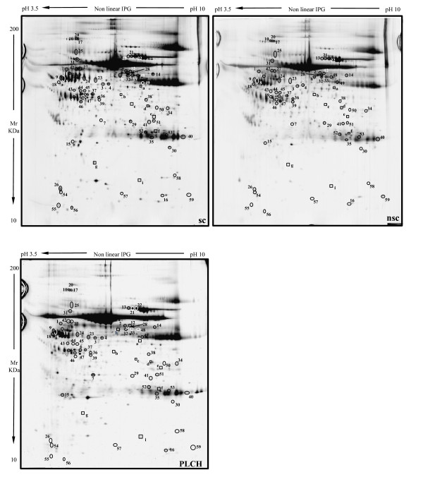
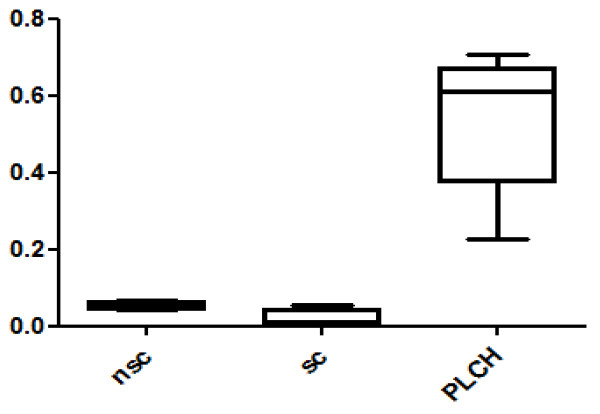
Results: Two-dimensional electrophoresis and image analysis revealed proteins that were differently expressed (quantitatively and qualitatively) in the three groups of subjects. The proteins were identified by mass spectrometry and have various functions (antioxidant, proinflammatory, antiprotease) and origins (plasma, locally produced, etc.). Many, such as protease inhibitors (human serpin B3) and antioxidant proteins (glutathione peroxidase and thioredoxin) are already linked to PLCH pathogenesis, whereas other proteins have never been associated with the disease. Interestingly, numerous proteolytic fragments of plasma proteins (including kininogen-1 N fragments and haptoglobin) were also identified and suggest increased proteolytic activity in this inflammatory lung disease. Differences in protein expression were found between the three groups and confirmed by Principal Component Analysis (PCA).
Conclusion: Analysis of BAL proteomes of PLCH patients and of smoker and non-smoker controls also proved to be useful for researching the pathogenetic mechanisms and for identifying biomarkers of this rare diffuse lung disease.

 求助内容:
求助内容: 应助结果提醒方式:


