{"title":"原发性胆汁性肝硬化中胆道上皮细胞凋亡、自噬和衰老。","authors":"Motoko Sasaki, Yasuni Nakanuma","doi":"10.1155/2010/205128","DOIUrl":null,"url":null,"abstract":"<p><p>Primary biliary cirrhosis (PBC) is a chronic cholestatic liver disease characterized serologically by the high prevalence of anti-mitochondrial autoantibodies (AMAs) and histologically by the cholangitis of small bile ducts, eventually followed by extensive loss of the small bile duct. An autoimmune pathogenesis is suggested by clinical and experimental studies, but there remain issues regarding the etiology, the significance of AMAs in the pathogenesis of bile duct lesions, and so on. The unique properties of apoptosis in biliary epithelial cells (BECs), in which there is exposure of autoantigen to the effectors of the immune system, are proposed to be a cause of bile duct lesions in PBC. Recent progress disclosed that cellular senescence and autophagy are involved in bile duct lesions in PBC. Senescent BECs may modulate the periductal microenvironment by expressing senescence-associated secretory phenotypes, including various chemokines, and contribute to the pathogenesis of bile duct lesions in PBC.</p>","PeriodicalId":73232,"journal":{"name":"Hepatitis research and treatment","volume":"2010 ","pages":"205128"},"PeriodicalIF":0.0000,"publicationDate":"2010-01-01","publicationTypes":"Journal Article","fieldsOfStudy":null,"isOpenAccess":false,"openAccessPdf":"https://sci-hub-pdf.com/10.1155/2010/205128","citationCount":"13","resultStr":"{\"title\":\"Biliary epithelial apoptosis, autophagy, and senescence in primary biliary cirrhosis.\",\"authors\":\"Motoko Sasaki, Yasuni Nakanuma\",\"doi\":\"10.1155/2010/205128\",\"DOIUrl\":null,\"url\":null,\"abstract\":\"<p><p>Primary biliary cirrhosis (PBC) is a chronic cholestatic liver disease characterized serologically by the high prevalence of anti-mitochondrial autoantibodies (AMAs) and histologically by the cholangitis of small bile ducts, eventually followed by extensive loss of the small bile duct. An autoimmune pathogenesis is suggested by clinical and experimental studies, but there remain issues regarding the etiology, the significance of AMAs in the pathogenesis of bile duct lesions, and so on. The unique properties of apoptosis in biliary epithelial cells (BECs), in which there is exposure of autoantigen to the effectors of the immune system, are proposed to be a cause of bile duct lesions in PBC. Recent progress disclosed that cellular senescence and autophagy are involved in bile duct lesions in PBC. Senescent BECs may modulate the periductal microenvironment by expressing senescence-associated secretory phenotypes, including various chemokines, and contribute to the pathogenesis of bile duct lesions in PBC.</p>\",\"PeriodicalId\":73232,\"journal\":{\"name\":\"Hepatitis research and treatment\",\"volume\":\"2010 \",\"pages\":\"205128\"},\"PeriodicalIF\":0.0000,\"publicationDate\":\"2010-01-01\",\"publicationTypes\":\"Journal Article\",\"fieldsOfStudy\":null,\"isOpenAccess\":false,\"openAccessPdf\":\"https://sci-hub-pdf.com/10.1155/2010/205128\",\"citationCount\":\"13\",\"resultStr\":null,\"platform\":\"Semanticscholar\",\"paperid\":null,\"PeriodicalName\":\"Hepatitis research and treatment\",\"FirstCategoryId\":\"1085\",\"ListUrlMain\":\"https://doi.org/10.1155/2010/205128\",\"RegionNum\":0,\"RegionCategory\":null,\"ArticlePicture\":[],\"TitleCN\":null,\"AbstractTextCN\":null,\"PMCID\":null,\"EPubDate\":\"2010/11/4 0:00:00\",\"PubModel\":\"Epub\",\"JCR\":\"\",\"JCRName\":\"\",\"Score\":null,\"Total\":0}","platform":"Semanticscholar","paperid":null,"PeriodicalName":"Hepatitis research and treatment","FirstCategoryId":"1085","ListUrlMain":"https://doi.org/10.1155/2010/205128","RegionNum":0,"RegionCategory":null,"ArticlePicture":[],"TitleCN":null,"AbstractTextCN":null,"PMCID":null,"EPubDate":"2010/11/4 0:00:00","PubModel":"Epub","JCR":"","JCRName":"","Score":null,"Total":0}
Biliary epithelial apoptosis, autophagy, and senescence in primary biliary cirrhosis.
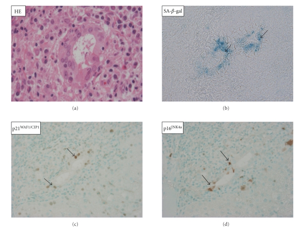
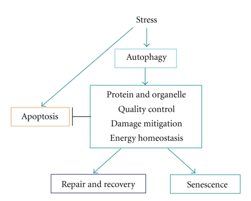
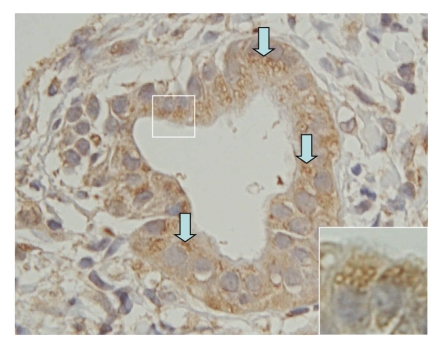
Primary biliary cirrhosis (PBC) is a chronic cholestatic liver disease characterized serologically by the high prevalence of anti-mitochondrial autoantibodies (AMAs) and histologically by the cholangitis of small bile ducts, eventually followed by extensive loss of the small bile duct. An autoimmune pathogenesis is suggested by clinical and experimental studies, but there remain issues regarding the etiology, the significance of AMAs in the pathogenesis of bile duct lesions, and so on. The unique properties of apoptosis in biliary epithelial cells (BECs), in which there is exposure of autoantigen to the effectors of the immune system, are proposed to be a cause of bile duct lesions in PBC. Recent progress disclosed that cellular senescence and autophagy are involved in bile duct lesions in PBC. Senescent BECs may modulate the periductal microenvironment by expressing senescence-associated secretory phenotypes, including various chemokines, and contribute to the pathogenesis of bile duct lesions in PBC.

 求助内容:
求助内容: 应助结果提醒方式:


