Aya Ishikawa, Motohiro Yamauchi, Keiji Suzuki, Shunichi Yamashita
{"title":"基于图像的DNA损伤信号定量测定揭示了电离辐射下G2检查点激活的阈值。","authors":"Aya Ishikawa, Motohiro Yamauchi, Keiji Suzuki, Shunichi Yamashita","doi":"10.1186/2041-9414-1-10","DOIUrl":null,"url":null,"abstract":"<p><strong>Background: </strong>Proteins involved in the DNA damage response accumulate as microscopically-visible nuclear foci on the chromatin flanking DNA double-strand breaks (DSBs). As growth of ionizing radiation (IR)-induced foci amplifies the ATM-dependent DNA damage signal, the formation of discrete foci plays a crucial role in cell cycle checkpoint activation, especially in cells exposed to lower doses of IR. However, there is no quantitative parameter for the foci which considers both the number and their size. Therefore, we have developed a novel parameter for DNA damage signal based on the image analysis of the foci and quantified the amount of the signal sufficient for G2 arrest.</p><p><strong>Results: </strong>The parameter that we have developed here was designated as SOID. SOID is an abbreviation of Sum Of Integrated Density, which represents the sum of fluorescence of each focus within one nucleus. The SOID was calculated for individual nucleus as the sum of (area (total pixel numbers) of each focus) x (mean fluorescence intensity per pixel of each focus). Therefore, the SOID accounts for the number, size, and fluorescence density of IR-induced foci, and the parameter reflects the flux of DNA damage signal much more accurately than foci number. Using very low doses of X-rays, we performed a \"two-way\" comparison of SOID of Ser139-phosphorylated histone H2AX foci between G2-arrested cells and mitosis-progressing cells, and between mitosis-progressing cells in the presence or absence of ATM or Chk1/2 inhibitor, both of which abrogate IR-induced G2/M checkpoint. The analysis revealed that there was a threshold of DNA damage signal for G2 arrest, which was around 4000~5000 SOID. G2 cells with < 4000 SOID were neglected by G2/M checkpoint, and thus, the cells could progress to mitosis. Chromosome analysis revealed that the checkpoint-neglected and mitosis-progressing cells had approximately two chromatid breaks on average, indicating that 4000~5000 SOID was equivalent to a few DNA double strand breaks.</p><p><strong>Conclusions: </strong>We developed a novel parameter for quantitative analysis of DNA damage signal, and we determined the threshold of DNA damage signal for IR-induced G2 arrest, which was represented by 4000~5000 SOID. The present study emphasizes that not only the foci number but also the size of the foci must be taken into consideration for the proper quantification of DNA damage signal.</p>","PeriodicalId":53596,"journal":{"name":"Genome Integrity","volume":"1 1","pages":"10"},"PeriodicalIF":0.0000,"publicationDate":"2010-08-04","publicationTypes":"Journal Article","fieldsOfStudy":null,"isOpenAccess":false,"openAccessPdf":"https://sci-hub-pdf.com/10.1186/2041-9414-1-10","citationCount":"13","resultStr":"{\"title\":\"Image-based quantitative determination of DNA damage signal reveals a threshold for G2 checkpoint activation in response to ionizing radiation.\",\"authors\":\"Aya Ishikawa, Motohiro Yamauchi, Keiji Suzuki, Shunichi Yamashita\",\"doi\":\"10.1186/2041-9414-1-10\",\"DOIUrl\":null,\"url\":null,\"abstract\":\"<p><strong>Background: </strong>Proteins involved in the DNA damage response accumulate as microscopically-visible nuclear foci on the chromatin flanking DNA double-strand breaks (DSBs). As growth of ionizing radiation (IR)-induced foci amplifies the ATM-dependent DNA damage signal, the formation of discrete foci plays a crucial role in cell cycle checkpoint activation, especially in cells exposed to lower doses of IR. However, there is no quantitative parameter for the foci which considers both the number and their size. Therefore, we have developed a novel parameter for DNA damage signal based on the image analysis of the foci and quantified the amount of the signal sufficient for G2 arrest.</p><p><strong>Results: </strong>The parameter that we have developed here was designated as SOID. SOID is an abbreviation of Sum Of Integrated Density, which represents the sum of fluorescence of each focus within one nucleus. The SOID was calculated for individual nucleus as the sum of (area (total pixel numbers) of each focus) x (mean fluorescence intensity per pixel of each focus). Therefore, the SOID accounts for the number, size, and fluorescence density of IR-induced foci, and the parameter reflects the flux of DNA damage signal much more accurately than foci number. Using very low doses of X-rays, we performed a \\\"two-way\\\" comparison of SOID of Ser139-phosphorylated histone H2AX foci between G2-arrested cells and mitosis-progressing cells, and between mitosis-progressing cells in the presence or absence of ATM or Chk1/2 inhibitor, both of which abrogate IR-induced G2/M checkpoint. The analysis revealed that there was a threshold of DNA damage signal for G2 arrest, which was around 4000~5000 SOID. G2 cells with < 4000 SOID were neglected by G2/M checkpoint, and thus, the cells could progress to mitosis. Chromosome analysis revealed that the checkpoint-neglected and mitosis-progressing cells had approximately two chromatid breaks on average, indicating that 4000~5000 SOID was equivalent to a few DNA double strand breaks.</p><p><strong>Conclusions: </strong>We developed a novel parameter for quantitative analysis of DNA damage signal, and we determined the threshold of DNA damage signal for IR-induced G2 arrest, which was represented by 4000~5000 SOID. The present study emphasizes that not only the foci number but also the size of the foci must be taken into consideration for the proper quantification of DNA damage signal.</p>\",\"PeriodicalId\":53596,\"journal\":{\"name\":\"Genome Integrity\",\"volume\":\"1 1\",\"pages\":\"10\"},\"PeriodicalIF\":0.0000,\"publicationDate\":\"2010-08-04\",\"publicationTypes\":\"Journal Article\",\"fieldsOfStudy\":null,\"isOpenAccess\":false,\"openAccessPdf\":\"https://sci-hub-pdf.com/10.1186/2041-9414-1-10\",\"citationCount\":\"13\",\"resultStr\":null,\"platform\":\"Semanticscholar\",\"paperid\":null,\"PeriodicalName\":\"Genome Integrity\",\"FirstCategoryId\":\"1085\",\"ListUrlMain\":\"https://doi.org/10.1186/2041-9414-1-10\",\"RegionNum\":0,\"RegionCategory\":null,\"ArticlePicture\":[],\"TitleCN\":null,\"AbstractTextCN\":null,\"PMCID\":null,\"EPubDate\":\"\",\"PubModel\":\"\",\"JCR\":\"Q4\",\"JCRName\":\"Biochemistry, Genetics and Molecular Biology\",\"Score\":null,\"Total\":0}","platform":"Semanticscholar","paperid":null,"PeriodicalName":"Genome Integrity","FirstCategoryId":"1085","ListUrlMain":"https://doi.org/10.1186/2041-9414-1-10","RegionNum":0,"RegionCategory":null,"ArticlePicture":[],"TitleCN":null,"AbstractTextCN":null,"PMCID":null,"EPubDate":"","PubModel":"","JCR":"Q4","JCRName":"Biochemistry, Genetics and Molecular Biology","Score":null,"Total":0}
引用次数: 13
摘要
背景:参与DNA损伤反应的蛋白质在染色质两侧DNA双链断裂(DSBs)上以显微镜可见的核焦点积累。由于电离辐射(IR)诱导的病灶的生长放大了atm依赖的DNA损伤信号,离散病灶的形成在细胞周期检查点激活中起着至关重要的作用,特别是在暴露于低剂量IR的细胞中。然而,没有一个定量的参数可以同时考虑到焦点的数量和大小。因此,我们基于对病灶的图像分析,开发了一种新的DNA损伤信号参数,并量化了足以引起G2阻滞的信号量。结果:我们建立的参数被指定为SOID。SOID是Sum of Integrated Density的缩写,它代表了一个原子核内每个焦点的荧光之和。单个细胞核的SOID计算为(每个焦点的面积(总像素数))x(每个焦点每像素的平均荧光强度)的总和。因此,SOID反映了红外诱导病灶的数量、大小和荧光密度,该参数比病灶数更准确地反映了DNA损伤信号的通量。使用非常低剂量的x射线,我们在G2阻滞细胞和有丝分裂进展细胞之间以及存在或不存在ATM或Chk1/2抑制剂的有丝分裂进展细胞之间进行了ser139磷酸化组蛋白H2AX灶的“双向”比较,这两种抑制剂都取消了ir诱导的G2/M检查点。分析发现,G2阻滞存在DNA损伤信号阈值,约为4000~5000 SOID。< 4000 SOID的G2细胞被G2/M检查点忽略,因此细胞可以进行有丝分裂。染色体分析显示,忽略检查点和有丝分裂进行的细胞平均约有2次染色单体断裂,表明4000~5000个SOID相当于少量DNA双链断裂。结论:我们建立了一个定量分析DNA损伤信号的新参数,并确定了ir诱导的G2阻滞的DNA损伤信号阈值,以4000~5000 SOID表示。本研究强调,要对DNA损伤信号进行定量,不仅要考虑病灶数,而且要考虑病灶的大小。
Image-based quantitative determination of DNA damage signal reveals a threshold for G2 checkpoint activation in response to ionizing radiation.
Background: Proteins involved in the DNA damage response accumulate as microscopically-visible nuclear foci on the chromatin flanking DNA double-strand breaks (DSBs). As growth of ionizing radiation (IR)-induced foci amplifies the ATM-dependent DNA damage signal, the formation of discrete foci plays a crucial role in cell cycle checkpoint activation, especially in cells exposed to lower doses of IR. However, there is no quantitative parameter for the foci which considers both the number and their size. Therefore, we have developed a novel parameter for DNA damage signal based on the image analysis of the foci and quantified the amount of the signal sufficient for G2 arrest.
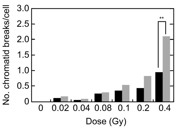
Results: The parameter that we have developed here was designated as SOID. SOID is an abbreviation of Sum Of Integrated Density, which represents the sum of fluorescence of each focus within one nucleus. The SOID was calculated for individual nucleus as the sum of (area (total pixel numbers) of each focus) x (mean fluorescence intensity per pixel of each focus). Therefore, the SOID accounts for the number, size, and fluorescence density of IR-induced foci, and the parameter reflects the flux of DNA damage signal much more accurately than foci number. Using very low doses of X-rays, we performed a "two-way" comparison of SOID of Ser139-phosphorylated histone H2AX foci between G2-arrested cells and mitosis-progressing cells, and between mitosis-progressing cells in the presence or absence of ATM or Chk1/2 inhibitor, both of which abrogate IR-induced G2/M checkpoint. The analysis revealed that there was a threshold of DNA damage signal for G2 arrest, which was around 4000~5000 SOID. G2 cells with < 4000 SOID were neglected by G2/M checkpoint, and thus, the cells could progress to mitosis. Chromosome analysis revealed that the checkpoint-neglected and mitosis-progressing cells had approximately two chromatid breaks on average, indicating that 4000~5000 SOID was equivalent to a few DNA double strand breaks.
Conclusions: We developed a novel parameter for quantitative analysis of DNA damage signal, and we determined the threshold of DNA damage signal for IR-induced G2 arrest, which was represented by 4000~5000 SOID. The present study emphasizes that not only the foci number but also the size of the foci must be taken into consideration for the proper quantification of DNA damage signal.

 求助内容:
求助内容: 应助结果提醒方式:


