{"title":"2D、3D高剂量、3D低剂量门控心肌82Rb PET显像比较。","authors":"Karin Knešaurek, Josef Machac, Jong Ho Kim","doi":"10.1186/1471-2385-7-4","DOIUrl":null,"url":null,"abstract":"<p><strong>Background: </strong>We compared 2D, 3D high dose (HD) and 3D low dose (LD) gated myocardial Rb-82 PET imaging in 16 normal human studies. The main goal in the paper is to evaluate whether the images obtained by a 3D LD studies are still of comparable clinical quality to the images obtained with the 2D HD or 3D HD studies.</p><p><strong>Methods: </strong>All 2D and 3D HD studies were performed with 2220 MBq of Rb-82. The 3D LD were performed with 740 MBq of Rb-82. A GE Advance PET system was used for acquisition. Polar maps were created and used to calculate noise among (NAS) and within (NWS) the segments in the noise analysis. In addition, the contrast between left ventricular (LV) wall and LV cavity was also analysed. For 13 subjects, ejection fraction (EF) on 2D and 3D studies was calculated using QGS program.</p><p><strong>Results: </strong>For the H20 reconstruction filter, the mean contrast in mid-ventricular short-axis slice was 0.33 +/- 0.06 for 2D studies. The same contrast for the 3D HD studies was 0.38 +/- 0.07 and for 3D LD, it was 0.34 +/- 0.08. For the 6 volunteers where 3D HD was used, NAS was 3.64*10-4 and NWS was 1.79*10-2 for 2D studies, and NAS was 3.70*10-4 and NWS was 1.85*10-2 for 3D HD studies, respectively. For the other 10 volunteers where 3D LD was used, NAS was 3.85*10-4 and NWS was 1.82*10-2 for the 2D studies, and NAS was 5.58*10-4 and NWS was 1.91*10-2 for the 3D LD studies, respectively. For the sharper H13 filter, the data followed the same pattern, with slightly higher values of contrast and noise. EF values in 2D and 3D were close. The Pearson's correlation coefficient was 0.90. The average difference from 13 subjects was 8.3%.</p><p><strong>Conclusion: </strong>2D and 3D HD gating Rb-82 PET cardiac studies have similar contrast, ejection fractions and noise levels. 3D LD gating imaging, gave comparable results in terms of contrast, EF and noise to either 2D or 3D HD gating PET imaging. 3D LD PET gated imaging can make Rb-82 PET cardiac imaging more affordable with significantly less radiation exposure to the patients.</p>","PeriodicalId":80684,"journal":{"name":"BMC nuclear medicine","volume":"7 ","pages":"4"},"PeriodicalIF":0.0000,"publicationDate":"2007-10-22","publicationTypes":"Journal Article","fieldsOfStudy":null,"isOpenAccess":false,"openAccessPdf":"https://sci-hub-pdf.com/10.1186/1471-2385-7-4","citationCount":"8","resultStr":"{\"title\":\"Comparison of 2D, 3D high dose and 3D low dose gated myocardial 82Rb PET imaging.\",\"authors\":\"Karin Knešaurek, Josef Machac, Jong Ho Kim\",\"doi\":\"10.1186/1471-2385-7-4\",\"DOIUrl\":null,\"url\":null,\"abstract\":\"<p><strong>Background: </strong>We compared 2D, 3D high dose (HD) and 3D low dose (LD) gated myocardial Rb-82 PET imaging in 16 normal human studies. The main goal in the paper is to evaluate whether the images obtained by a 3D LD studies are still of comparable clinical quality to the images obtained with the 2D HD or 3D HD studies.</p><p><strong>Methods: </strong>All 2D and 3D HD studies were performed with 2220 MBq of Rb-82. The 3D LD were performed with 740 MBq of Rb-82. A GE Advance PET system was used for acquisition. Polar maps were created and used to calculate noise among (NAS) and within (NWS) the segments in the noise analysis. In addition, the contrast between left ventricular (LV) wall and LV cavity was also analysed. For 13 subjects, ejection fraction (EF) on 2D and 3D studies was calculated using QGS program.</p><p><strong>Results: </strong>For the H20 reconstruction filter, the mean contrast in mid-ventricular short-axis slice was 0.33 +/- 0.06 for 2D studies. The same contrast for the 3D HD studies was 0.38 +/- 0.07 and for 3D LD, it was 0.34 +/- 0.08. For the 6 volunteers where 3D HD was used, NAS was 3.64*10-4 and NWS was 1.79*10-2 for 2D studies, and NAS was 3.70*10-4 and NWS was 1.85*10-2 for 3D HD studies, respectively. For the other 10 volunteers where 3D LD was used, NAS was 3.85*10-4 and NWS was 1.82*10-2 for the 2D studies, and NAS was 5.58*10-4 and NWS was 1.91*10-2 for the 3D LD studies, respectively. For the sharper H13 filter, the data followed the same pattern, with slightly higher values of contrast and noise. EF values in 2D and 3D were close. The Pearson's correlation coefficient was 0.90. The average difference from 13 subjects was 8.3%.</p><p><strong>Conclusion: </strong>2D and 3D HD gating Rb-82 PET cardiac studies have similar contrast, ejection fractions and noise levels. 3D LD gating imaging, gave comparable results in terms of contrast, EF and noise to either 2D or 3D HD gating PET imaging. 3D LD PET gated imaging can make Rb-82 PET cardiac imaging more affordable with significantly less radiation exposure to the patients.</p>\",\"PeriodicalId\":80684,\"journal\":{\"name\":\"BMC nuclear medicine\",\"volume\":\"7 \",\"pages\":\"4\"},\"PeriodicalIF\":0.0000,\"publicationDate\":\"2007-10-22\",\"publicationTypes\":\"Journal Article\",\"fieldsOfStudy\":null,\"isOpenAccess\":false,\"openAccessPdf\":\"https://sci-hub-pdf.com/10.1186/1471-2385-7-4\",\"citationCount\":\"8\",\"resultStr\":null,\"platform\":\"Semanticscholar\",\"paperid\":null,\"PeriodicalName\":\"BMC nuclear medicine\",\"FirstCategoryId\":\"1085\",\"ListUrlMain\":\"https://doi.org/10.1186/1471-2385-7-4\",\"RegionNum\":0,\"RegionCategory\":null,\"ArticlePicture\":[],\"TitleCN\":null,\"AbstractTextCN\":null,\"PMCID\":null,\"EPubDate\":\"\",\"PubModel\":\"\",\"JCR\":\"\",\"JCRName\":\"\",\"Score\":null,\"Total\":0}","platform":"Semanticscholar","paperid":null,"PeriodicalName":"BMC nuclear medicine","FirstCategoryId":"1085","ListUrlMain":"https://doi.org/10.1186/1471-2385-7-4","RegionNum":0,"RegionCategory":null,"ArticlePicture":[],"TitleCN":null,"AbstractTextCN":null,"PMCID":null,"EPubDate":"","PubModel":"","JCR":"","JCRName":"","Score":null,"Total":0}
Comparison of 2D, 3D high dose and 3D low dose gated myocardial 82Rb PET imaging.
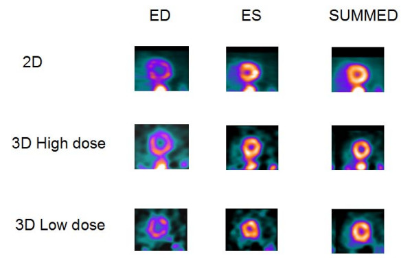
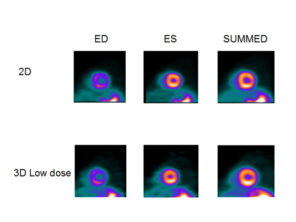
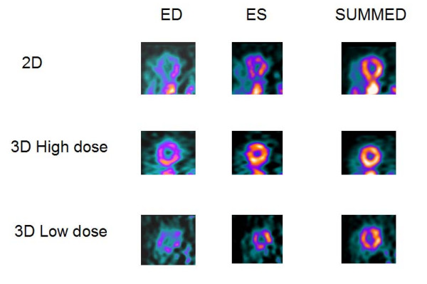
Background: We compared 2D, 3D high dose (HD) and 3D low dose (LD) gated myocardial Rb-82 PET imaging in 16 normal human studies. The main goal in the paper is to evaluate whether the images obtained by a 3D LD studies are still of comparable clinical quality to the images obtained with the 2D HD or 3D HD studies.
Methods: All 2D and 3D HD studies were performed with 2220 MBq of Rb-82. The 3D LD were performed with 740 MBq of Rb-82. A GE Advance PET system was used for acquisition. Polar maps were created and used to calculate noise among (NAS) and within (NWS) the segments in the noise analysis. In addition, the contrast between left ventricular (LV) wall and LV cavity was also analysed. For 13 subjects, ejection fraction (EF) on 2D and 3D studies was calculated using QGS program.
Results: For the H20 reconstruction filter, the mean contrast in mid-ventricular short-axis slice was 0.33 +/- 0.06 for 2D studies. The same contrast for the 3D HD studies was 0.38 +/- 0.07 and for 3D LD, it was 0.34 +/- 0.08. For the 6 volunteers where 3D HD was used, NAS was 3.64*10-4 and NWS was 1.79*10-2 for 2D studies, and NAS was 3.70*10-4 and NWS was 1.85*10-2 for 3D HD studies, respectively. For the other 10 volunteers where 3D LD was used, NAS was 3.85*10-4 and NWS was 1.82*10-2 for the 2D studies, and NAS was 5.58*10-4 and NWS was 1.91*10-2 for the 3D LD studies, respectively. For the sharper H13 filter, the data followed the same pattern, with slightly higher values of contrast and noise. EF values in 2D and 3D were close. The Pearson's correlation coefficient was 0.90. The average difference from 13 subjects was 8.3%.
Conclusion: 2D and 3D HD gating Rb-82 PET cardiac studies have similar contrast, ejection fractions and noise levels. 3D LD gating imaging, gave comparable results in terms of contrast, EF and noise to either 2D or 3D HD gating PET imaging. 3D LD PET gated imaging can make Rb-82 PET cardiac imaging more affordable with significantly less radiation exposure to the patients.

 求助内容:
求助内容: 应助结果提醒方式:


