{"title":"18F SPECT与PET在心肌成像中的比较:一个真实的胸-心虚研究。","authors":"Karin Knešaurek, Josef Machac","doi":"10.1186/1471-2385-6-5","DOIUrl":null,"url":null,"abstract":"<p><strong>Background: </strong>Positron emission tomography (PET) imaging with fluorine-18 (18F) Fluorodeoxyglucose (FDG) and flow tracer such as Rubidium-82 (82Rb) is an established method for evaluating an ischemic but viable myocardium. However, the high cost of PET imaging restricts its wider clinical use. Therefore, less expensive 18F FDG single photon emission computed tomography (SPECT) imaging has been considered as an alternative to 18F FDG PET imaging. The purpose of the work is to compare SPECT with PET in myocardial perfusion/viability imaging.</p><p><strong>Methods: </strong>A nonuniform RH-2 thorax-heart phantom was used in the SPECT and PET acquisitions. Three inserts, 3 cm, 2 cm and 1 cm in diameter, were placed in the left ventricular (LV) wall to simulate infarcts. The phantom acquisition was performed sequentially with 7.4 MBq of 18F and 22.2 MBq of Technetium-99m (99mTc) in the SPECT study and with 7.4 MBq of 18F and 370 MBq of 82Rb in the PET study. SPECT and PET data were processed using standard reconstruction software provided by vendors. Circumferential profiles of the short-axis slices, the contrast and viability of the inserts were used to evaluate the SPECT and PET images.</p><p><strong>Results: </strong>The contrast for 3 cm, 2 cm and 1 cm inserts were for 18F PET data, 1.0 +/- 0.01, 0.67 +/- 0.02 and 0.25 +/- 0.01, respectively. For 82Rb PET data, the corresponding contrast values were 0.61 +/- 0.02, 0.37 +/- 0.02 and 0.19 +/- 0.01, respectively. For 18F SPECT the contrast values were, 0.31 +/- 0.03 and 0.20 +/- 0.05 for 3 cm and 2 cm inserts, respectively. For 99mTc SPECT the contrast values were, 0.63 +/- 0.04 and 0.24 +/- 0.05 for 3 cm and 2 cm inserts respectively. In SPECT, the 1 cm insert was not detectable. In the SPECT study, all three inserts were falsely diagnosed as \"viable\", while in the PET study, only the 1 cm insert was diagnosed falsely \"viable\".</p><p><strong>Conclusion: </strong>For smaller defects the 99mTc/18F SPECT imaging cannot entirely replace the more expensive 82Rb/18F PET for myocardial perfusion/viability imaging, due to poorer image spatial resolution and poorer defect contrast.</p>","PeriodicalId":80684,"journal":{"name":"BMC nuclear medicine","volume":"6 ","pages":"5"},"PeriodicalIF":0.0000,"publicationDate":"2006-10-31","publicationTypes":"Journal Article","fieldsOfStudy":null,"isOpenAccess":false,"openAccessPdf":"https://sci-hub-pdf.com/10.1186/1471-2385-6-5","citationCount":"14","resultStr":"{\"title\":\"Comparison of 18F SPECT with PET in myocardial imaging: a realistic thorax-cardiac phantom study.\",\"authors\":\"Karin Knešaurek, Josef Machac\",\"doi\":\"10.1186/1471-2385-6-5\",\"DOIUrl\":null,\"url\":null,\"abstract\":\"<p><strong>Background: </strong>Positron emission tomography (PET) imaging with fluorine-18 (18F) Fluorodeoxyglucose (FDG) and flow tracer such as Rubidium-82 (82Rb) is an established method for evaluating an ischemic but viable myocardium. However, the high cost of PET imaging restricts its wider clinical use. Therefore, less expensive 18F FDG single photon emission computed tomography (SPECT) imaging has been considered as an alternative to 18F FDG PET imaging. The purpose of the work is to compare SPECT with PET in myocardial perfusion/viability imaging.</p><p><strong>Methods: </strong>A nonuniform RH-2 thorax-heart phantom was used in the SPECT and PET acquisitions. Three inserts, 3 cm, 2 cm and 1 cm in diameter, were placed in the left ventricular (LV) wall to simulate infarcts. The phantom acquisition was performed sequentially with 7.4 MBq of 18F and 22.2 MBq of Technetium-99m (99mTc) in the SPECT study and with 7.4 MBq of 18F and 370 MBq of 82Rb in the PET study. SPECT and PET data were processed using standard reconstruction software provided by vendors. Circumferential profiles of the short-axis slices, the contrast and viability of the inserts were used to evaluate the SPECT and PET images.</p><p><strong>Results: </strong>The contrast for 3 cm, 2 cm and 1 cm inserts were for 18F PET data, 1.0 +/- 0.01, 0.67 +/- 0.02 and 0.25 +/- 0.01, respectively. For 82Rb PET data, the corresponding contrast values were 0.61 +/- 0.02, 0.37 +/- 0.02 and 0.19 +/- 0.01, respectively. For 18F SPECT the contrast values were, 0.31 +/- 0.03 and 0.20 +/- 0.05 for 3 cm and 2 cm inserts, respectively. For 99mTc SPECT the contrast values were, 0.63 +/- 0.04 and 0.24 +/- 0.05 for 3 cm and 2 cm inserts respectively. In SPECT, the 1 cm insert was not detectable. In the SPECT study, all three inserts were falsely diagnosed as \\\"viable\\\", while in the PET study, only the 1 cm insert was diagnosed falsely \\\"viable\\\".</p><p><strong>Conclusion: </strong>For smaller defects the 99mTc/18F SPECT imaging cannot entirely replace the more expensive 82Rb/18F PET for myocardial perfusion/viability imaging, due to poorer image spatial resolution and poorer defect contrast.</p>\",\"PeriodicalId\":80684,\"journal\":{\"name\":\"BMC nuclear medicine\",\"volume\":\"6 \",\"pages\":\"5\"},\"PeriodicalIF\":0.0000,\"publicationDate\":\"2006-10-31\",\"publicationTypes\":\"Journal Article\",\"fieldsOfStudy\":null,\"isOpenAccess\":false,\"openAccessPdf\":\"https://sci-hub-pdf.com/10.1186/1471-2385-6-5\",\"citationCount\":\"14\",\"resultStr\":null,\"platform\":\"Semanticscholar\",\"paperid\":null,\"PeriodicalName\":\"BMC nuclear medicine\",\"FirstCategoryId\":\"1085\",\"ListUrlMain\":\"https://doi.org/10.1186/1471-2385-6-5\",\"RegionNum\":0,\"RegionCategory\":null,\"ArticlePicture\":[],\"TitleCN\":null,\"AbstractTextCN\":null,\"PMCID\":null,\"EPubDate\":\"\",\"PubModel\":\"\",\"JCR\":\"\",\"JCRName\":\"\",\"Score\":null,\"Total\":0}","platform":"Semanticscholar","paperid":null,"PeriodicalName":"BMC nuclear medicine","FirstCategoryId":"1085","ListUrlMain":"https://doi.org/10.1186/1471-2385-6-5","RegionNum":0,"RegionCategory":null,"ArticlePicture":[],"TitleCN":null,"AbstractTextCN":null,"PMCID":null,"EPubDate":"","PubModel":"","JCR":"","JCRName":"","Score":null,"Total":0}
Comparison of 18F SPECT with PET in myocardial imaging: a realistic thorax-cardiac phantom study.
Background: Positron emission tomography (PET) imaging with fluorine-18 (18F) Fluorodeoxyglucose (FDG) and flow tracer such as Rubidium-82 (82Rb) is an established method for evaluating an ischemic but viable myocardium. However, the high cost of PET imaging restricts its wider clinical use. Therefore, less expensive 18F FDG single photon emission computed tomography (SPECT) imaging has been considered as an alternative to 18F FDG PET imaging. The purpose of the work is to compare SPECT with PET in myocardial perfusion/viability imaging.
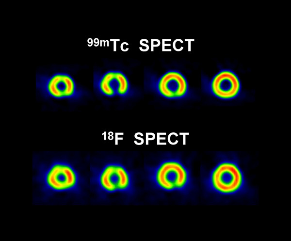
Methods: A nonuniform RH-2 thorax-heart phantom was used in the SPECT and PET acquisitions. Three inserts, 3 cm, 2 cm and 1 cm in diameter, were placed in the left ventricular (LV) wall to simulate infarcts. The phantom acquisition was performed sequentially with 7.4 MBq of 18F and 22.2 MBq of Technetium-99m (99mTc) in the SPECT study and with 7.4 MBq of 18F and 370 MBq of 82Rb in the PET study. SPECT and PET data were processed using standard reconstruction software provided by vendors. Circumferential profiles of the short-axis slices, the contrast and viability of the inserts were used to evaluate the SPECT and PET images.
Results: The contrast for 3 cm, 2 cm and 1 cm inserts were for 18F PET data, 1.0 +/- 0.01, 0.67 +/- 0.02 and 0.25 +/- 0.01, respectively. For 82Rb PET data, the corresponding contrast values were 0.61 +/- 0.02, 0.37 +/- 0.02 and 0.19 +/- 0.01, respectively. For 18F SPECT the contrast values were, 0.31 +/- 0.03 and 0.20 +/- 0.05 for 3 cm and 2 cm inserts, respectively. For 99mTc SPECT the contrast values were, 0.63 +/- 0.04 and 0.24 +/- 0.05 for 3 cm and 2 cm inserts respectively. In SPECT, the 1 cm insert was not detectable. In the SPECT study, all three inserts were falsely diagnosed as "viable", while in the PET study, only the 1 cm insert was diagnosed falsely "viable".
Conclusion: For smaller defects the 99mTc/18F SPECT imaging cannot entirely replace the more expensive 82Rb/18F PET for myocardial perfusion/viability imaging, due to poorer image spatial resolution and poorer defect contrast.


 求助内容:
求助内容: 应助结果提醒方式:


