{"title":"正常和烧伤皮肤中人体防御素的免疫荧光反褶积显微镜和图像重建。","authors":"Brian J Poindexter","doi":"","DOIUrl":null,"url":null,"abstract":"<p><strong>Objective: </strong>The aim of this study was visualization and localization of the human antimicrobials human beta defensins 1, 2, and 3, neutrophil defensin alpha (human neutrophil peptide), and the cathelicidin LL-37 in normal and burned skin, and determination of the cell types in which these antimicrobials were localized.</p><p><strong>Methods: </strong>Tissue sections were probed with antimicrobial antibodies, tagged with fluorescently labeled secondary antibodies, and subjected to fluorescence deconvolution microscopy and image reconstruction. Images were generated by stacking multiple-section scans, which were then volume rendered by rotating stacks 360 degrees about an axis, or modeled in 3 dimensions.</p><p><strong>Results: </strong>This technique yields a definitive image, providing a rapid basis for further quantification and manipulation from a full 3-dimensional aspect. In normal skin, human beta defensin-1 was localized to the perinuclear region of keratinocytes; human beta defensin-2 was primarily localized to the stratum germinativum; human beta defensin-3 was found in dendritic cells of the stratum spinosum; human neutrophil peptide was randomly distributed in the papillary dermis; and LL-37 was concentrated in the stratum corneum and along ducts. In burned skin, in which keratinocytes are lost or destroyed, human beta defensin-1 was present in dermal glandular structures including hair shafts; human beta defensin-2 and human beta defensin-3 were found in the remaining keratin layers and glands of the lower dermis; human neutrophil peptide was primarily localized to hair shafts, though visible in residual keratin layers; and LL-37 was evident in very high concentrations in the epithelium of sweat ducts.</p><p><strong>Conclusion: </strong>We conclude via this technique that cells in the lower dermal and subdermal regions of burned skin synthesize antimicrobials after burn injury, and maintain something of a barrier against infection. This methodology is discussed and explained in this article.</p>","PeriodicalId":87438,"journal":{"name":"Journal of burns and wounds","volume":null,"pages":null},"PeriodicalIF":0.0000,"publicationDate":"2005-04-25","publicationTypes":"Journal Article","fieldsOfStudy":null,"isOpenAccess":false,"openAccessPdf":"https://www.ncbi.nlm.nih.gov/pmc/articles/PMC1501114/pdf/","citationCount":"0","resultStr":"{\"title\":\"Immunofluorescence deconvolution microscopy and image reconstruction of human defensins in normal and burned skin.\",\"authors\":\"Brian J Poindexter\",\"doi\":\"\",\"DOIUrl\":null,\"url\":null,\"abstract\":\"<p><strong>Objective: </strong>The aim of this study was visualization and localization of the human antimicrobials human beta defensins 1, 2, and 3, neutrophil defensin alpha (human neutrophil peptide), and the cathelicidin LL-37 in normal and burned skin, and determination of the cell types in which these antimicrobials were localized.</p><p><strong>Methods: </strong>Tissue sections were probed with antimicrobial antibodies, tagged with fluorescently labeled secondary antibodies, and subjected to fluorescence deconvolution microscopy and image reconstruction. Images were generated by stacking multiple-section scans, which were then volume rendered by rotating stacks 360 degrees about an axis, or modeled in 3 dimensions.</p><p><strong>Results: </strong>This technique yields a definitive image, providing a rapid basis for further quantification and manipulation from a full 3-dimensional aspect. In normal skin, human beta defensin-1 was localized to the perinuclear region of keratinocytes; human beta defensin-2 was primarily localized to the stratum germinativum; human beta defensin-3 was found in dendritic cells of the stratum spinosum; human neutrophil peptide was randomly distributed in the papillary dermis; and LL-37 was concentrated in the stratum corneum and along ducts. In burned skin, in which keratinocytes are lost or destroyed, human beta defensin-1 was present in dermal glandular structures including hair shafts; human beta defensin-2 and human beta defensin-3 were found in the remaining keratin layers and glands of the lower dermis; human neutrophil peptide was primarily localized to hair shafts, though visible in residual keratin layers; and LL-37 was evident in very high concentrations in the epithelium of sweat ducts.</p><p><strong>Conclusion: </strong>We conclude via this technique that cells in the lower dermal and subdermal regions of burned skin synthesize antimicrobials after burn injury, and maintain something of a barrier against infection. This methodology is discussed and explained in this article.</p>\",\"PeriodicalId\":87438,\"journal\":{\"name\":\"Journal of burns and wounds\",\"volume\":null,\"pages\":null},\"PeriodicalIF\":0.0000,\"publicationDate\":\"2005-04-25\",\"publicationTypes\":\"Journal Article\",\"fieldsOfStudy\":null,\"isOpenAccess\":false,\"openAccessPdf\":\"https://www.ncbi.nlm.nih.gov/pmc/articles/PMC1501114/pdf/\",\"citationCount\":\"0\",\"resultStr\":null,\"platform\":\"Semanticscholar\",\"paperid\":null,\"PeriodicalName\":\"Journal of burns and wounds\",\"FirstCategoryId\":\"1085\",\"ListUrlMain\":\"\",\"RegionNum\":0,\"RegionCategory\":null,\"ArticlePicture\":[],\"TitleCN\":null,\"AbstractTextCN\":null,\"PMCID\":null,\"EPubDate\":\"\",\"PubModel\":\"\",\"JCR\":\"\",\"JCRName\":\"\",\"Score\":null,\"Total\":0}","platform":"Semanticscholar","paperid":null,"PeriodicalName":"Journal of burns and wounds","FirstCategoryId":"1085","ListUrlMain":"","RegionNum":0,"RegionCategory":null,"ArticlePicture":[],"TitleCN":null,"AbstractTextCN":null,"PMCID":null,"EPubDate":"","PubModel":"","JCR":"","JCRName":"","Score":null,"Total":0}
Immunofluorescence deconvolution microscopy and image reconstruction of human defensins in normal and burned skin.
Objective: The aim of this study was visualization and localization of the human antimicrobials human beta defensins 1, 2, and 3, neutrophil defensin alpha (human neutrophil peptide), and the cathelicidin LL-37 in normal and burned skin, and determination of the cell types in which these antimicrobials were localized.
Methods: Tissue sections were probed with antimicrobial antibodies, tagged with fluorescently labeled secondary antibodies, and subjected to fluorescence deconvolution microscopy and image reconstruction. Images were generated by stacking multiple-section scans, which were then volume rendered by rotating stacks 360 degrees about an axis, or modeled in 3 dimensions.
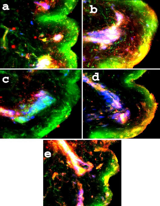
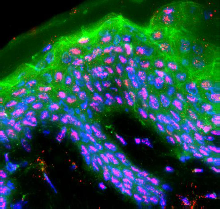
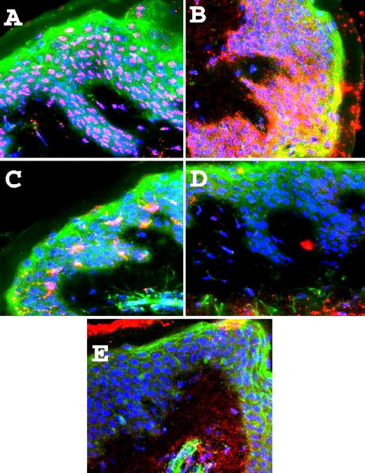
Results: This technique yields a definitive image, providing a rapid basis for further quantification and manipulation from a full 3-dimensional aspect. In normal skin, human beta defensin-1 was localized to the perinuclear region of keratinocytes; human beta defensin-2 was primarily localized to the stratum germinativum; human beta defensin-3 was found in dendritic cells of the stratum spinosum; human neutrophil peptide was randomly distributed in the papillary dermis; and LL-37 was concentrated in the stratum corneum and along ducts. In burned skin, in which keratinocytes are lost or destroyed, human beta defensin-1 was present in dermal glandular structures including hair shafts; human beta defensin-2 and human beta defensin-3 were found in the remaining keratin layers and glands of the lower dermis; human neutrophil peptide was primarily localized to hair shafts, though visible in residual keratin layers; and LL-37 was evident in very high concentrations in the epithelium of sweat ducts.
Conclusion: We conclude via this technique that cells in the lower dermal and subdermal regions of burned skin synthesize antimicrobials after burn injury, and maintain something of a barrier against infection. This methodology is discussed and explained in this article.

 求助内容:
求助内容: 应助结果提醒方式:


