Adhish Basu, Sarath Chandra Sistla, Surendra Kumar Verma, S Jagdish
{"title":"腋窝淋巴肿大——一种不寻常的丝虫病表现。","authors":"Adhish Basu, Sarath Chandra Sistla, Surendra Kumar Verma, S Jagdish","doi":"10.1186/1475-2883-5-9","DOIUrl":null,"url":null,"abstract":"<p><p>Clinical manifestations of lymphatic filariasis depend on the area of lymphatic involvement and the duration of infection. A 21 year old man, resident in a filariasis endemic region, presented with multiple matted lymph nodes with cystic areas forming a large mass in his left axilla. An ultrasound scan of the axilla using a 7.5 MHz transducer revealed grossly dilated lymphatics but no filarial dance sign. Fine needle (21 G) aspiration cytology (FNAC) from the dilated lymphatics and solid areas in the lymph node mass revealed multiple microfilariae in a background of reactive lymphoid cells. Peripheral blood smears revealed microfilaremia with significant eosinophilia. Diagnosis of left axillary Bancroftian lymphadenovarix was made. On the administration of oral diethylcarbamazine, the diameter of the lymphatic vessels in the lymphadenovarix reduced considerably in size and microfilaremia disappeared. We report this case because axillary lymphadenovarix is a rare presentation of filariasis. This case is also unique since microfilariae were demonstrated in the fluid aspirated from the dilated lymphatics of the lymphadenovarix in the absence of live adult worms.</p>","PeriodicalId":84756,"journal":{"name":"Filaria journal","volume":"5 ","pages":"9"},"PeriodicalIF":0.0000,"publicationDate":"2006-07-30","publicationTypes":"Journal Article","fieldsOfStudy":null,"isOpenAccess":false,"openAccessPdf":"https://sci-hub-pdf.com/10.1186/1475-2883-5-9","citationCount":"21","resultStr":"{\"title\":\"Lymphadenovarix in the axilla--an unusual presentation of filariasis.\",\"authors\":\"Adhish Basu, Sarath Chandra Sistla, Surendra Kumar Verma, S Jagdish\",\"doi\":\"10.1186/1475-2883-5-9\",\"DOIUrl\":null,\"url\":null,\"abstract\":\"<p><p>Clinical manifestations of lymphatic filariasis depend on the area of lymphatic involvement and the duration of infection. A 21 year old man, resident in a filariasis endemic region, presented with multiple matted lymph nodes with cystic areas forming a large mass in his left axilla. An ultrasound scan of the axilla using a 7.5 MHz transducer revealed grossly dilated lymphatics but no filarial dance sign. Fine needle (21 G) aspiration cytology (FNAC) from the dilated lymphatics and solid areas in the lymph node mass revealed multiple microfilariae in a background of reactive lymphoid cells. Peripheral blood smears revealed microfilaremia with significant eosinophilia. Diagnosis of left axillary Bancroftian lymphadenovarix was made. On the administration of oral diethylcarbamazine, the diameter of the lymphatic vessels in the lymphadenovarix reduced considerably in size and microfilaremia disappeared. We report this case because axillary lymphadenovarix is a rare presentation of filariasis. This case is also unique since microfilariae were demonstrated in the fluid aspirated from the dilated lymphatics of the lymphadenovarix in the absence of live adult worms.</p>\",\"PeriodicalId\":84756,\"journal\":{\"name\":\"Filaria journal\",\"volume\":\"5 \",\"pages\":\"9\"},\"PeriodicalIF\":0.0000,\"publicationDate\":\"2006-07-30\",\"publicationTypes\":\"Journal Article\",\"fieldsOfStudy\":null,\"isOpenAccess\":false,\"openAccessPdf\":\"https://sci-hub-pdf.com/10.1186/1475-2883-5-9\",\"citationCount\":\"21\",\"resultStr\":null,\"platform\":\"Semanticscholar\",\"paperid\":null,\"PeriodicalName\":\"Filaria journal\",\"FirstCategoryId\":\"1085\",\"ListUrlMain\":\"https://doi.org/10.1186/1475-2883-5-9\",\"RegionNum\":0,\"RegionCategory\":null,\"ArticlePicture\":[],\"TitleCN\":null,\"AbstractTextCN\":null,\"PMCID\":null,\"EPubDate\":\"\",\"PubModel\":\"\",\"JCR\":\"\",\"JCRName\":\"\",\"Score\":null,\"Total\":0}","platform":"Semanticscholar","paperid":null,"PeriodicalName":"Filaria journal","FirstCategoryId":"1085","ListUrlMain":"https://doi.org/10.1186/1475-2883-5-9","RegionNum":0,"RegionCategory":null,"ArticlePicture":[],"TitleCN":null,"AbstractTextCN":null,"PMCID":null,"EPubDate":"","PubModel":"","JCR":"","JCRName":"","Score":null,"Total":0}
Lymphadenovarix in the axilla--an unusual presentation of filariasis.
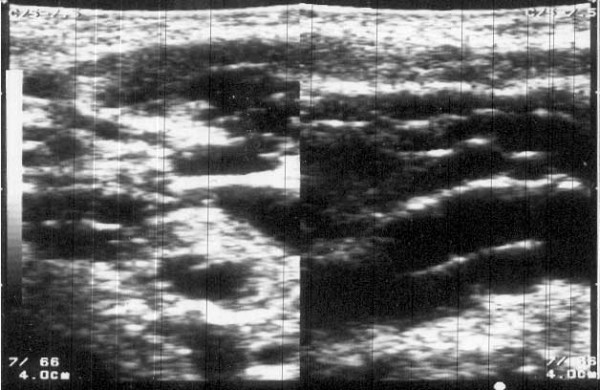
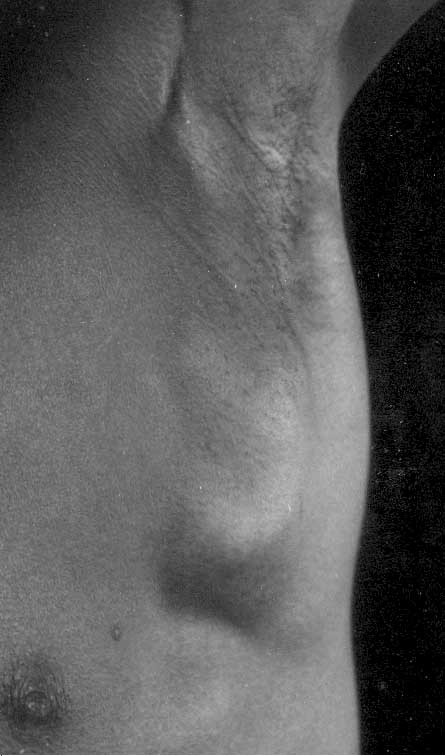
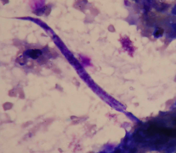
Clinical manifestations of lymphatic filariasis depend on the area of lymphatic involvement and the duration of infection. A 21 year old man, resident in a filariasis endemic region, presented with multiple matted lymph nodes with cystic areas forming a large mass in his left axilla. An ultrasound scan of the axilla using a 7.5 MHz transducer revealed grossly dilated lymphatics but no filarial dance sign. Fine needle (21 G) aspiration cytology (FNAC) from the dilated lymphatics and solid areas in the lymph node mass revealed multiple microfilariae in a background of reactive lymphoid cells. Peripheral blood smears revealed microfilaremia with significant eosinophilia. Diagnosis of left axillary Bancroftian lymphadenovarix was made. On the administration of oral diethylcarbamazine, the diameter of the lymphatic vessels in the lymphadenovarix reduced considerably in size and microfilaremia disappeared. We report this case because axillary lymphadenovarix is a rare presentation of filariasis. This case is also unique since microfilariae were demonstrated in the fluid aspirated from the dilated lymphatics of the lymphadenovarix in the absence of live adult worms.

 求助内容:
求助内容: 应助结果提醒方式:


