Daya R Pokharel, Reeta Rai, Pankaj Kumar, C M Chaturvedi, Sushma Rathaur
{"title":"牛丝虫子宫颈蛇尾虫胶原酶和亮氨酸氨基肽酶的组织定位。","authors":"Daya R Pokharel, Reeta Rai, Pankaj Kumar, C M Chaturvedi, Sushma Rathaur","doi":"10.1186/1475-2883-5-7","DOIUrl":null,"url":null,"abstract":"<p><strong>Background: </strong>Like other helminth proteases, filarial proteases have also been shown to require for parasite survival inside the host and mediate various physiologic processes such as tissue invasion, feeding, embryogenesis and host immune evasion. Many of these proteases have shown potential for vaccines and chemotherapeutic agents against active filarial infections. Setaria cervi is a bovine filarial parasite and serves as a good parasite model for the studies in lymphatic filariasis. Recently, a 175 kDa collagenase and leucine aminopeptidase (LAP) have been purified and characterized from the bovine filarial parasite S. cervi and shown to be potential vaccine candidate and diagnostic marker, respectively for human lymphatic filariasis. However, their tissue localizations and putative roles in the parasite biology have not yet been examined and thus remain unclear. Therefore, the current study attempts to localize and explore the putative roles of these two enzymes in S. cervi.</p><p><strong>Methods: </strong>The tissue distributions of 175 kDa collagenase and leucine aminopeptidase in S. cervi were examined by immunohistochemical and histochemical methods, respectively. Immune sera obtained from the jirds immunized with collagenase served as primary antibody, rabbit anti-mouse IgG-HRP conjugate as secondary antibody and DAB as the substrate for the immunostaining of collagenase. Leu-betaNA was used as the substrate for the histochemical staining of LAP.</p><p><strong>Results: </strong>Both the collagenase and LAP were present in the body wall; however, they differ in their distribution pattern in different layers of body wall. Collagenase was mainly localized in epicuticle, cuticle, syncytial hypodermis and the nerve cord region whereas LAP was more concentrated in epicuticle, longitudinal muscle layers and almost absent or very faintly stained in syncytial hypodermis and nerve cord region. Both collagenase and LAP showed their common distributions in intestine, uterus and mature eggs, growing embryos and mf. Very strong immunostaining of collagenase in the outer body surface of the parasite indicates its major role in host-parasite relationship whereas the presence of LAP in muscular region suggests its role in tissue remodeling. The common presences of collagenase and LAP in the S. cervi intestine, ovary, uterus, eggs and mf suggest that they also have collaborative roles in molting, nutrition and embryogenesis. The data obtained on their immunological characterizations and their presence in important parasite organs give strong indication that they are critical for the survival of filarial parasite and thus can be good vaccine candidates and/or diagnostic markers for human lymphatic filariasis.</p><p><strong>Conclusion: </strong>The manuscript reports for the first time the tissue distribution of collagenase and LAP in the bovine filarial parasite S. cervi and discuss their putative roles in vivo. Our findings also open the avenue to examine the roles of these two proteases in vivo, which will require further experiments like using their natural substrates and/or specific inhibitors in each tissues.</p>","PeriodicalId":84756,"journal":{"name":"Filaria journal","volume":"5 ","pages":"7"},"PeriodicalIF":0.0000,"publicationDate":"2006-05-22","publicationTypes":"Journal Article","fieldsOfStudy":null,"isOpenAccess":false,"openAccessPdf":"https://sci-hub-pdf.com/10.1186/1475-2883-5-7","citationCount":"16","resultStr":"{\"title\":\"Tissue localization of collagenase and leucine aminopeptidase in the bovine filarial parasite Setaria cervi.\",\"authors\":\"Daya R Pokharel, Reeta Rai, Pankaj Kumar, C M Chaturvedi, Sushma Rathaur\",\"doi\":\"10.1186/1475-2883-5-7\",\"DOIUrl\":null,\"url\":null,\"abstract\":\"<p><strong>Background: </strong>Like other helminth proteases, filarial proteases have also been shown to require for parasite survival inside the host and mediate various physiologic processes such as tissue invasion, feeding, embryogenesis and host immune evasion. Many of these proteases have shown potential for vaccines and chemotherapeutic agents against active filarial infections. Setaria cervi is a bovine filarial parasite and serves as a good parasite model for the studies in lymphatic filariasis. Recently, a 175 kDa collagenase and leucine aminopeptidase (LAP) have been purified and characterized from the bovine filarial parasite S. cervi and shown to be potential vaccine candidate and diagnostic marker, respectively for human lymphatic filariasis. However, their tissue localizations and putative roles in the parasite biology have not yet been examined and thus remain unclear. Therefore, the current study attempts to localize and explore the putative roles of these two enzymes in S. cervi.</p><p><strong>Methods: </strong>The tissue distributions of 175 kDa collagenase and leucine aminopeptidase in S. cervi were examined by immunohistochemical and histochemical methods, respectively. Immune sera obtained from the jirds immunized with collagenase served as primary antibody, rabbit anti-mouse IgG-HRP conjugate as secondary antibody and DAB as the substrate for the immunostaining of collagenase. Leu-betaNA was used as the substrate for the histochemical staining of LAP.</p><p><strong>Results: </strong>Both the collagenase and LAP were present in the body wall; however, they differ in their distribution pattern in different layers of body wall. Collagenase was mainly localized in epicuticle, cuticle, syncytial hypodermis and the nerve cord region whereas LAP was more concentrated in epicuticle, longitudinal muscle layers and almost absent or very faintly stained in syncytial hypodermis and nerve cord region. Both collagenase and LAP showed their common distributions in intestine, uterus and mature eggs, growing embryos and mf. Very strong immunostaining of collagenase in the outer body surface of the parasite indicates its major role in host-parasite relationship whereas the presence of LAP in muscular region suggests its role in tissue remodeling. The common presences of collagenase and LAP in the S. cervi intestine, ovary, uterus, eggs and mf suggest that they also have collaborative roles in molting, nutrition and embryogenesis. The data obtained on their immunological characterizations and their presence in important parasite organs give strong indication that they are critical for the survival of filarial parasite and thus can be good vaccine candidates and/or diagnostic markers for human lymphatic filariasis.</p><p><strong>Conclusion: </strong>The manuscript reports for the first time the tissue distribution of collagenase and LAP in the bovine filarial parasite S. cervi and discuss their putative roles in vivo. Our findings also open the avenue to examine the roles of these two proteases in vivo, which will require further experiments like using their natural substrates and/or specific inhibitors in each tissues.</p>\",\"PeriodicalId\":84756,\"journal\":{\"name\":\"Filaria journal\",\"volume\":\"5 \",\"pages\":\"7\"},\"PeriodicalIF\":0.0000,\"publicationDate\":\"2006-05-22\",\"publicationTypes\":\"Journal Article\",\"fieldsOfStudy\":null,\"isOpenAccess\":false,\"openAccessPdf\":\"https://sci-hub-pdf.com/10.1186/1475-2883-5-7\",\"citationCount\":\"16\",\"resultStr\":null,\"platform\":\"Semanticscholar\",\"paperid\":null,\"PeriodicalName\":\"Filaria journal\",\"FirstCategoryId\":\"1085\",\"ListUrlMain\":\"https://doi.org/10.1186/1475-2883-5-7\",\"RegionNum\":0,\"RegionCategory\":null,\"ArticlePicture\":[],\"TitleCN\":null,\"AbstractTextCN\":null,\"PMCID\":null,\"EPubDate\":\"\",\"PubModel\":\"\",\"JCR\":\"\",\"JCRName\":\"\",\"Score\":null,\"Total\":0}","platform":"Semanticscholar","paperid":null,"PeriodicalName":"Filaria journal","FirstCategoryId":"1085","ListUrlMain":"https://doi.org/10.1186/1475-2883-5-7","RegionNum":0,"RegionCategory":null,"ArticlePicture":[],"TitleCN":null,"AbstractTextCN":null,"PMCID":null,"EPubDate":"","PubModel":"","JCR":"","JCRName":"","Score":null,"Total":0}
Tissue localization of collagenase and leucine aminopeptidase in the bovine filarial parasite Setaria cervi.
Background: Like other helminth proteases, filarial proteases have also been shown to require for parasite survival inside the host and mediate various physiologic processes such as tissue invasion, feeding, embryogenesis and host immune evasion. Many of these proteases have shown potential for vaccines and chemotherapeutic agents against active filarial infections. Setaria cervi is a bovine filarial parasite and serves as a good parasite model for the studies in lymphatic filariasis. Recently, a 175 kDa collagenase and leucine aminopeptidase (LAP) have been purified and characterized from the bovine filarial parasite S. cervi and shown to be potential vaccine candidate and diagnostic marker, respectively for human lymphatic filariasis. However, their tissue localizations and putative roles in the parasite biology have not yet been examined and thus remain unclear. Therefore, the current study attempts to localize and explore the putative roles of these two enzymes in S. cervi.
Methods: The tissue distributions of 175 kDa collagenase and leucine aminopeptidase in S. cervi were examined by immunohistochemical and histochemical methods, respectively. Immune sera obtained from the jirds immunized with collagenase served as primary antibody, rabbit anti-mouse IgG-HRP conjugate as secondary antibody and DAB as the substrate for the immunostaining of collagenase. Leu-betaNA was used as the substrate for the histochemical staining of LAP.
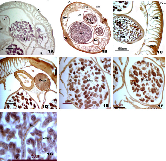
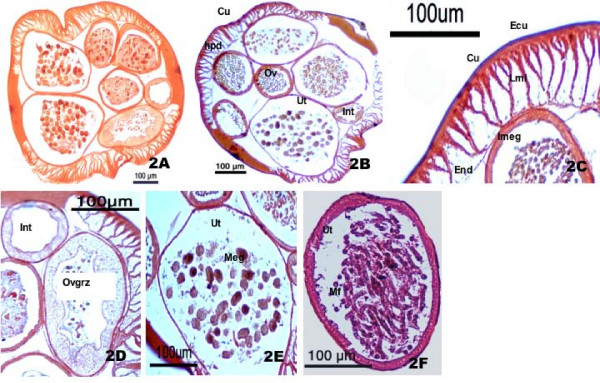
Results: Both the collagenase and LAP were present in the body wall; however, they differ in their distribution pattern in different layers of body wall. Collagenase was mainly localized in epicuticle, cuticle, syncytial hypodermis and the nerve cord region whereas LAP was more concentrated in epicuticle, longitudinal muscle layers and almost absent or very faintly stained in syncytial hypodermis and nerve cord region. Both collagenase and LAP showed their common distributions in intestine, uterus and mature eggs, growing embryos and mf. Very strong immunostaining of collagenase in the outer body surface of the parasite indicates its major role in host-parasite relationship whereas the presence of LAP in muscular region suggests its role in tissue remodeling. The common presences of collagenase and LAP in the S. cervi intestine, ovary, uterus, eggs and mf suggest that they also have collaborative roles in molting, nutrition and embryogenesis. The data obtained on their immunological characterizations and their presence in important parasite organs give strong indication that they are critical for the survival of filarial parasite and thus can be good vaccine candidates and/or diagnostic markers for human lymphatic filariasis.
Conclusion: The manuscript reports for the first time the tissue distribution of collagenase and LAP in the bovine filarial parasite S. cervi and discuss their putative roles in vivo. Our findings also open the avenue to examine the roles of these two proteases in vivo, which will require further experiments like using their natural substrates and/or specific inhibitors in each tissues.

 求助内容:
求助内容: 应助结果提醒方式:


