Teresa Massardo, Omar Alonso, Augusto Llamas-Ollier, Levin Kabasakal, Uma Ravishankar, Rossana Morales, Lucía Delgado, Ajit K Padhy
{"title":"一项国际多中心试验的结果表明,在乳腺癌方案的腋窝评估中,应谨慎考虑平面Tc99m- sestamibi扫描造影术。","authors":"Teresa Massardo, Omar Alonso, Augusto Llamas-Ollier, Levin Kabasakal, Uma Ravishankar, Rossana Morales, Lucía Delgado, Ajit K Padhy","doi":"10.1186/1471-2385-5-4","DOIUrl":null,"url":null,"abstract":"<p><strong>Background: </strong>Lymph node status is the most important prognostic indicator in breast cancer in recently diagnosed primary lesion. As a part of an interregional protocol using scintimammography with Tc99m compounds, the value of planar Tc99m sestamibi scanning for axillary lymph node evaluation is presented. Since there is a wide range of reported values, a standardized protocol of planar imaging was performed.</p><p><strong>Methods: </strong>One hundred and forty-nine female patients were included prospectively from different regions. Their mean age was 55.1 +/- 11.9 years. Histological report was obtained from 2.987 excised lymph nodes from 150 axillas. An early planar chest image was obtained at 10 min in all patients and a delayed one in 95 patients, all images performed with 740-925 MBq dose of Tc99m sestamibi. Blind lecture of all axillary regions was interpreted by 2 independent observers considering any well defined focal area of increased uptake as an involved axilla. Diagnostic values, 95% confidence intervals [CI] and also likelihood ratios (LR) were calculated.</p><p><strong>Results: </strong>Node histology demonstrated tumor involvement in 546 out of 2987 lymph nodes. Sestamibi was positive in 30 axillas (25 true-positive) and negative in 120 (only 55 true-negative). The sensitivity corresponded to 27.8% [CI = 18.9-38.2] and specificity to 91.7% [81.6-97.2]. The positive and negative LR were 3.33 and 0.79, respectively. There was no difference between early and delayed images. Sensitivity was higher in patients with palpable lesions.</p><p><strong>Conclusion: </strong>This work confirmed that non tomographic Tc99m sestamibi scintimammography had a very low detection rate for axillary lymph node involvement and it should not be applied for clinical assessment of breast cancer.</p>","PeriodicalId":80684,"journal":{"name":"BMC nuclear medicine","volume":"5 ","pages":"4"},"PeriodicalIF":0.0000,"publicationDate":"2005-07-27","publicationTypes":"Journal Article","fieldsOfStudy":null,"isOpenAccess":false,"openAccessPdf":"https://sci-hub-pdf.com/10.1186/1471-2385-5-4","citationCount":"12","resultStr":"{\"title\":\"Planar Tc99m--sestamibi scintimammography should be considered cautiously in the axillary evaluation of breast cancer protocols: results of an international multicenter trial.\",\"authors\":\"Teresa Massardo, Omar Alonso, Augusto Llamas-Ollier, Levin Kabasakal, Uma Ravishankar, Rossana Morales, Lucía Delgado, Ajit K Padhy\",\"doi\":\"10.1186/1471-2385-5-4\",\"DOIUrl\":null,\"url\":null,\"abstract\":\"<p><strong>Background: </strong>Lymph node status is the most important prognostic indicator in breast cancer in recently diagnosed primary lesion. As a part of an interregional protocol using scintimammography with Tc99m compounds, the value of planar Tc99m sestamibi scanning for axillary lymph node evaluation is presented. Since there is a wide range of reported values, a standardized protocol of planar imaging was performed.</p><p><strong>Methods: </strong>One hundred and forty-nine female patients were included prospectively from different regions. Their mean age was 55.1 +/- 11.9 years. Histological report was obtained from 2.987 excised lymph nodes from 150 axillas. An early planar chest image was obtained at 10 min in all patients and a delayed one in 95 patients, all images performed with 740-925 MBq dose of Tc99m sestamibi. Blind lecture of all axillary regions was interpreted by 2 independent observers considering any well defined focal area of increased uptake as an involved axilla. Diagnostic values, 95% confidence intervals [CI] and also likelihood ratios (LR) were calculated.</p><p><strong>Results: </strong>Node histology demonstrated tumor involvement in 546 out of 2987 lymph nodes. Sestamibi was positive in 30 axillas (25 true-positive) and negative in 120 (only 55 true-negative). The sensitivity corresponded to 27.8% [CI = 18.9-38.2] and specificity to 91.7% [81.6-97.2]. The positive and negative LR were 3.33 and 0.79, respectively. There was no difference between early and delayed images. Sensitivity was higher in patients with palpable lesions.</p><p><strong>Conclusion: </strong>This work confirmed that non tomographic Tc99m sestamibi scintimammography had a very low detection rate for axillary lymph node involvement and it should not be applied for clinical assessment of breast cancer.</p>\",\"PeriodicalId\":80684,\"journal\":{\"name\":\"BMC nuclear medicine\",\"volume\":\"5 \",\"pages\":\"4\"},\"PeriodicalIF\":0.0000,\"publicationDate\":\"2005-07-27\",\"publicationTypes\":\"Journal Article\",\"fieldsOfStudy\":null,\"isOpenAccess\":false,\"openAccessPdf\":\"https://sci-hub-pdf.com/10.1186/1471-2385-5-4\",\"citationCount\":\"12\",\"resultStr\":null,\"platform\":\"Semanticscholar\",\"paperid\":null,\"PeriodicalName\":\"BMC nuclear medicine\",\"FirstCategoryId\":\"1085\",\"ListUrlMain\":\"https://doi.org/10.1186/1471-2385-5-4\",\"RegionNum\":0,\"RegionCategory\":null,\"ArticlePicture\":[],\"TitleCN\":null,\"AbstractTextCN\":null,\"PMCID\":null,\"EPubDate\":\"\",\"PubModel\":\"\",\"JCR\":\"\",\"JCRName\":\"\",\"Score\":null,\"Total\":0}","platform":"Semanticscholar","paperid":null,"PeriodicalName":"BMC nuclear medicine","FirstCategoryId":"1085","ListUrlMain":"https://doi.org/10.1186/1471-2385-5-4","RegionNum":0,"RegionCategory":null,"ArticlePicture":[],"TitleCN":null,"AbstractTextCN":null,"PMCID":null,"EPubDate":"","PubModel":"","JCR":"","JCRName":"","Score":null,"Total":0}
Planar Tc99m--sestamibi scintimammography should be considered cautiously in the axillary evaluation of breast cancer protocols: results of an international multicenter trial.
Background: Lymph node status is the most important prognostic indicator in breast cancer in recently diagnosed primary lesion. As a part of an interregional protocol using scintimammography with Tc99m compounds, the value of planar Tc99m sestamibi scanning for axillary lymph node evaluation is presented. Since there is a wide range of reported values, a standardized protocol of planar imaging was performed.
Methods: One hundred and forty-nine female patients were included prospectively from different regions. Their mean age was 55.1 +/- 11.9 years. Histological report was obtained from 2.987 excised lymph nodes from 150 axillas. An early planar chest image was obtained at 10 min in all patients and a delayed one in 95 patients, all images performed with 740-925 MBq dose of Tc99m sestamibi. Blind lecture of all axillary regions was interpreted by 2 independent observers considering any well defined focal area of increased uptake as an involved axilla. Diagnostic values, 95% confidence intervals [CI] and also likelihood ratios (LR) were calculated.
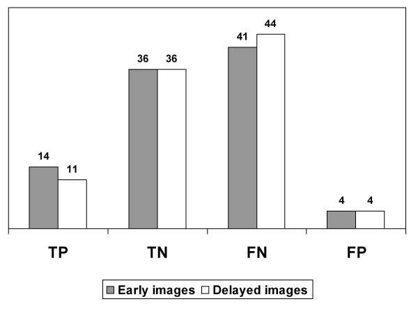
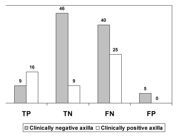
Results: Node histology demonstrated tumor involvement in 546 out of 2987 lymph nodes. Sestamibi was positive in 30 axillas (25 true-positive) and negative in 120 (only 55 true-negative). The sensitivity corresponded to 27.8% [CI = 18.9-38.2] and specificity to 91.7% [81.6-97.2]. The positive and negative LR were 3.33 and 0.79, respectively. There was no difference between early and delayed images. Sensitivity was higher in patients with palpable lesions.
Conclusion: This work confirmed that non tomographic Tc99m sestamibi scintimammography had a very low detection rate for axillary lymph node involvement and it should not be applied for clinical assessment of breast cancer.

 求助内容:
求助内容: 应助结果提醒方式:


