{"title":"ERK活性传感器的研制,一种体外基于fret的细胞外调节激酶活性传感器。","authors":"Harry M Green, José Alberola-Ila","doi":"10.1186/1472-6769-5-1","DOIUrl":null,"url":null,"abstract":"<p><strong>Background: </strong>Study of ERK activation has thus far relied on biochemical assays that are limited to the use of phospho-specific antibodies and radioactivity in vitro, and analysis of whole cell populations in vivo. As with many systems, fluorescence resonance energy transfer (FRET) can be utilized to make highly sensitive detectors of molecular activity. Here we introduce FRET-based ERK Activity Sensors, which utilize variants of Enhanced Green Fluorescent Protein fused by an ERK-specific peptide linker to detect ERK2 activity.</p><p><strong>Results: </strong>ERK Activity Sensors display varying changes in FRET upon phosphorylation by active ERK2 in vitro depending on the composition of ERK-specific peptide linker sequences derived from known in vivo ERK targets, Ets1 and Elk1. Analysis of point mutations reveals specific residues involved in ERK binding and phosphorylation of ERK Activity Sensor 3. ERK2 also shows high in vitro specificity for these sensors over two other major MAP Kinases, p38 and pSAPK/JNK.</p><p><strong>Conclusion: </strong>EAS's are a convenient, non-radioactive alternative to study ERK dynamics in vitro. They can be utilized to study ERK activity in real-time. This new technology can be applied to studying ERK kinetics in vitro, analysis of ERK activity in whole cell extracts, and high-throughput screening technologies.</p>","PeriodicalId":80682,"journal":{"name":"BMC chemical biology","volume":"5 ","pages":"1"},"PeriodicalIF":0.0000,"publicationDate":"2005-07-05","publicationTypes":"Journal Article","fieldsOfStudy":null,"isOpenAccess":false,"openAccessPdf":"https://www.ncbi.nlm.nih.gov/pmc/articles/PMC1180429/pdf/","citationCount":"0","resultStr":"{\"title\":\"Development of ERK Activity Sensor, an in vitro, FRET-based sensor of Extracellular Regulated Kinase activity.\",\"authors\":\"Harry M Green, José Alberola-Ila\",\"doi\":\"10.1186/1472-6769-5-1\",\"DOIUrl\":null,\"url\":null,\"abstract\":\"<p><strong>Background: </strong>Study of ERK activation has thus far relied on biochemical assays that are limited to the use of phospho-specific antibodies and radioactivity in vitro, and analysis of whole cell populations in vivo. As with many systems, fluorescence resonance energy transfer (FRET) can be utilized to make highly sensitive detectors of molecular activity. Here we introduce FRET-based ERK Activity Sensors, which utilize variants of Enhanced Green Fluorescent Protein fused by an ERK-specific peptide linker to detect ERK2 activity.</p><p><strong>Results: </strong>ERK Activity Sensors display varying changes in FRET upon phosphorylation by active ERK2 in vitro depending on the composition of ERK-specific peptide linker sequences derived from known in vivo ERK targets, Ets1 and Elk1. Analysis of point mutations reveals specific residues involved in ERK binding and phosphorylation of ERK Activity Sensor 3. ERK2 also shows high in vitro specificity for these sensors over two other major MAP Kinases, p38 and pSAPK/JNK.</p><p><strong>Conclusion: </strong>EAS's are a convenient, non-radioactive alternative to study ERK dynamics in vitro. They can be utilized to study ERK activity in real-time. This new technology can be applied to studying ERK kinetics in vitro, analysis of ERK activity in whole cell extracts, and high-throughput screening technologies.</p>\",\"PeriodicalId\":80682,\"journal\":{\"name\":\"BMC chemical biology\",\"volume\":\"5 \",\"pages\":\"1\"},\"PeriodicalIF\":0.0000,\"publicationDate\":\"2005-07-05\",\"publicationTypes\":\"Journal Article\",\"fieldsOfStudy\":null,\"isOpenAccess\":false,\"openAccessPdf\":\"https://www.ncbi.nlm.nih.gov/pmc/articles/PMC1180429/pdf/\",\"citationCount\":\"0\",\"resultStr\":null,\"platform\":\"Semanticscholar\",\"paperid\":null,\"PeriodicalName\":\"BMC chemical biology\",\"FirstCategoryId\":\"1085\",\"ListUrlMain\":\"https://doi.org/10.1186/1472-6769-5-1\",\"RegionNum\":0,\"RegionCategory\":null,\"ArticlePicture\":[],\"TitleCN\":null,\"AbstractTextCN\":null,\"PMCID\":null,\"EPubDate\":\"\",\"PubModel\":\"\",\"JCR\":\"\",\"JCRName\":\"\",\"Score\":null,\"Total\":0}","platform":"Semanticscholar","paperid":null,"PeriodicalName":"BMC chemical biology","FirstCategoryId":"1085","ListUrlMain":"https://doi.org/10.1186/1472-6769-5-1","RegionNum":0,"RegionCategory":null,"ArticlePicture":[],"TitleCN":null,"AbstractTextCN":null,"PMCID":null,"EPubDate":"","PubModel":"","JCR":"","JCRName":"","Score":null,"Total":0}
Development of ERK Activity Sensor, an in vitro, FRET-based sensor of Extracellular Regulated Kinase activity.
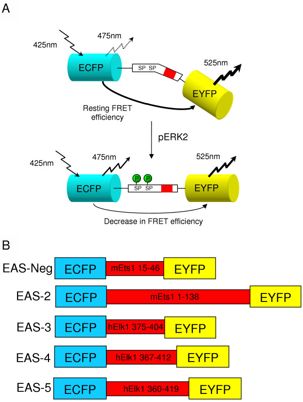
Background: Study of ERK activation has thus far relied on biochemical assays that are limited to the use of phospho-specific antibodies and radioactivity in vitro, and analysis of whole cell populations in vivo. As with many systems, fluorescence resonance energy transfer (FRET) can be utilized to make highly sensitive detectors of molecular activity. Here we introduce FRET-based ERK Activity Sensors, which utilize variants of Enhanced Green Fluorescent Protein fused by an ERK-specific peptide linker to detect ERK2 activity.
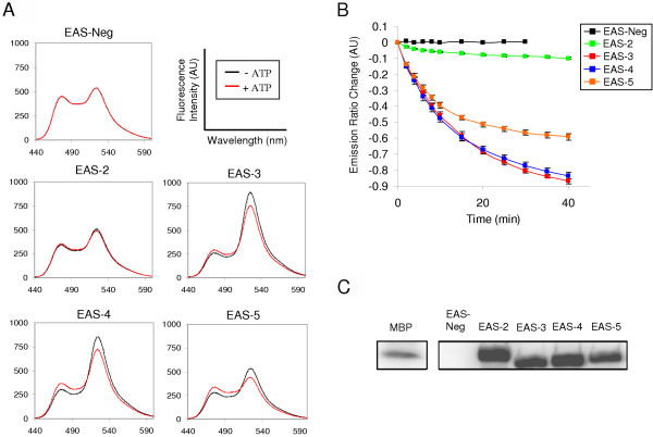
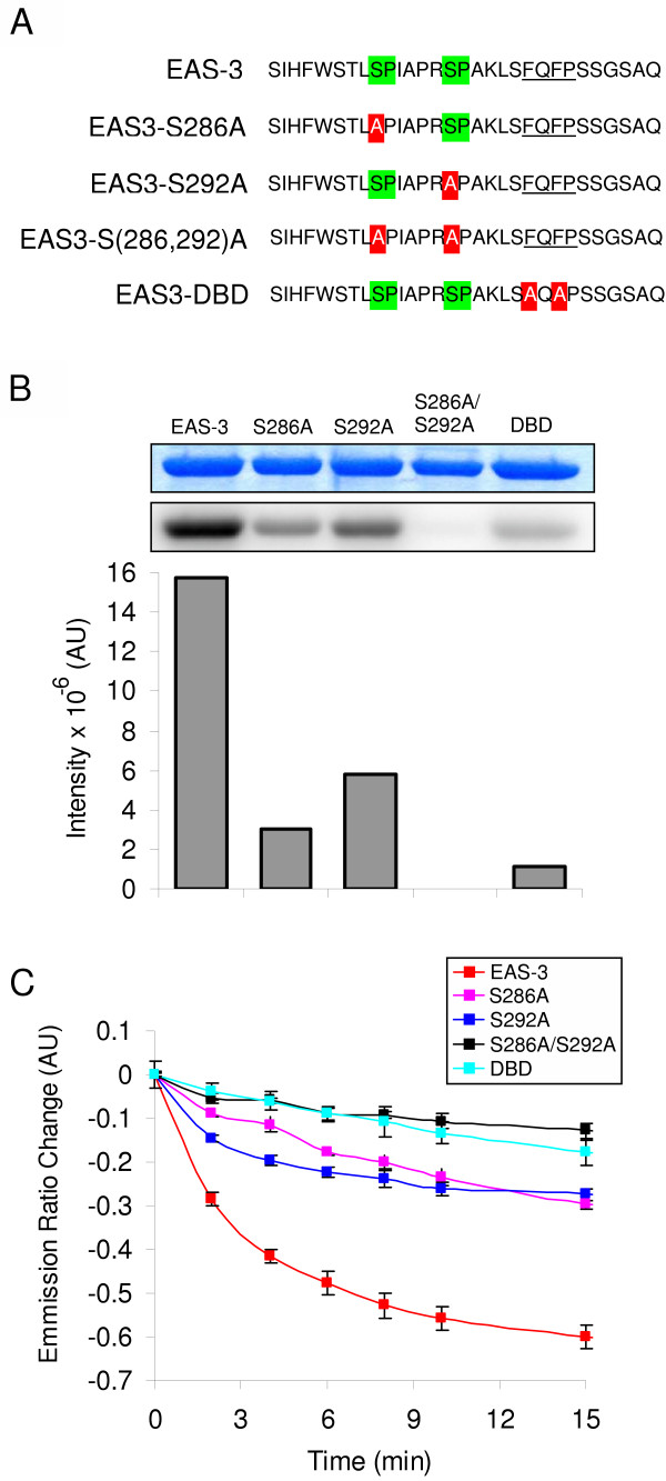
Results: ERK Activity Sensors display varying changes in FRET upon phosphorylation by active ERK2 in vitro depending on the composition of ERK-specific peptide linker sequences derived from known in vivo ERK targets, Ets1 and Elk1. Analysis of point mutations reveals specific residues involved in ERK binding and phosphorylation of ERK Activity Sensor 3. ERK2 also shows high in vitro specificity for these sensors over two other major MAP Kinases, p38 and pSAPK/JNK.
Conclusion: EAS's are a convenient, non-radioactive alternative to study ERK dynamics in vitro. They can be utilized to study ERK activity in real-time. This new technology can be applied to studying ERK kinetics in vitro, analysis of ERK activity in whole cell extracts, and high-throughput screening technologies.

 求助内容:
求助内容: 应助结果提醒方式:


