Shuang Xue , Zhoufeng Zhang , Siyuan Li , Jian Du , Hang Zhao , Meijie Qi , Chenglong Tao
{"title":"术中甲状腺病变高光谱融合与纯特征提取框架","authors":"Shuang Xue , Zhoufeng Zhang , Siyuan Li , Jian Du , Hang Zhao , Meijie Qi , Chenglong Tao","doi":"10.1016/j.media.2025.103832","DOIUrl":null,"url":null,"abstract":"<div><div>Thyroid cancer has remained one of the most prevalent endocrine malignancies. In routine surgery, thyroid cancer analysis involves two time-consuming steps: intraoperative frozen section preparation and manual microscopic examination. Recently, info-rich hyperspectral intelligence analysis has been studied, reducing subjective bias but only optimizing the intraoperative second step and the model complexity, ignoring the independent features that possess substance fingerprints. To bridge the gaps, we developed a hyperspectral recognition algorithm called PS4EM-SN for intraoperatively ex-vivo macro thyroid lesion, which comprised a pure spectral with pure spatial(SPS) learning framework and a spatial–spectral fusion embed mechanism(SSEM) coupled with cascade attention. The cascade attention mechanism, integrating Squeeze-and-Excitation (SE) and Non-Local (NOL) blocks, enhanced robustness to the outliers of SSEM and improved generalization. The experimental results were satisfactory in differentiating non-malignant and malignant regions with 93.91% average accuracy. Given its hyperspectral multifaceted performance, our method promises a digital solution for intraoperative thyroid diagnosis.</div></div>","PeriodicalId":18328,"journal":{"name":"Medical image analysis","volume":"107 ","pages":"Article 103832"},"PeriodicalIF":11.8000,"publicationDate":"2025-10-06","publicationTypes":"Journal Article","fieldsOfStudy":null,"isOpenAccess":false,"openAccessPdf":"","citationCount":"0","resultStr":"{\"title\":\"Fusion and pure feature extraction framework for intraoperative hyperspectral of thyroid lesion\",\"authors\":\"Shuang Xue , Zhoufeng Zhang , Siyuan Li , Jian Du , Hang Zhao , Meijie Qi , Chenglong Tao\",\"doi\":\"10.1016/j.media.2025.103832\",\"DOIUrl\":null,\"url\":null,\"abstract\":\"<div><div>Thyroid cancer has remained one of the most prevalent endocrine malignancies. In routine surgery, thyroid cancer analysis involves two time-consuming steps: intraoperative frozen section preparation and manual microscopic examination. Recently, info-rich hyperspectral intelligence analysis has been studied, reducing subjective bias but only optimizing the intraoperative second step and the model complexity, ignoring the independent features that possess substance fingerprints. To bridge the gaps, we developed a hyperspectral recognition algorithm called PS4EM-SN for intraoperatively ex-vivo macro thyroid lesion, which comprised a pure spectral with pure spatial(SPS) learning framework and a spatial–spectral fusion embed mechanism(SSEM) coupled with cascade attention. The cascade attention mechanism, integrating Squeeze-and-Excitation (SE) and Non-Local (NOL) blocks, enhanced robustness to the outliers of SSEM and improved generalization. The experimental results were satisfactory in differentiating non-malignant and malignant regions with 93.91% average accuracy. Given its hyperspectral multifaceted performance, our method promises a digital solution for intraoperative thyroid diagnosis.</div></div>\",\"PeriodicalId\":18328,\"journal\":{\"name\":\"Medical image analysis\",\"volume\":\"107 \",\"pages\":\"Article 103832\"},\"PeriodicalIF\":11.8000,\"publicationDate\":\"2025-10-06\",\"publicationTypes\":\"Journal Article\",\"fieldsOfStudy\":null,\"isOpenAccess\":false,\"openAccessPdf\":\"\",\"citationCount\":\"0\",\"resultStr\":null,\"platform\":\"Semanticscholar\",\"paperid\":null,\"PeriodicalName\":\"Medical image analysis\",\"FirstCategoryId\":\"5\",\"ListUrlMain\":\"https://www.sciencedirect.com/science/article/pii/S1361841525003780\",\"RegionNum\":1,\"RegionCategory\":\"医学\",\"ArticlePicture\":[],\"TitleCN\":null,\"AbstractTextCN\":null,\"PMCID\":null,\"EPubDate\":\"\",\"PubModel\":\"\",\"JCR\":\"Q1\",\"JCRName\":\"COMPUTER SCIENCE, ARTIFICIAL INTELLIGENCE\",\"Score\":null,\"Total\":0}","platform":"Semanticscholar","paperid":null,"PeriodicalName":"Medical image analysis","FirstCategoryId":"5","ListUrlMain":"https://www.sciencedirect.com/science/article/pii/S1361841525003780","RegionNum":1,"RegionCategory":"医学","ArticlePicture":[],"TitleCN":null,"AbstractTextCN":null,"PMCID":null,"EPubDate":"","PubModel":"","JCR":"Q1","JCRName":"COMPUTER SCIENCE, ARTIFICIAL INTELLIGENCE","Score":null,"Total":0}
Fusion and pure feature extraction framework for intraoperative hyperspectral of thyroid lesion
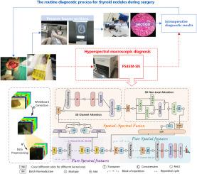
Thyroid cancer has remained one of the most prevalent endocrine malignancies. In routine surgery, thyroid cancer analysis involves two time-consuming steps: intraoperative frozen section preparation and manual microscopic examination. Recently, info-rich hyperspectral intelligence analysis has been studied, reducing subjective bias but only optimizing the intraoperative second step and the model complexity, ignoring the independent features that possess substance fingerprints. To bridge the gaps, we developed a hyperspectral recognition algorithm called PS4EM-SN for intraoperatively ex-vivo macro thyroid lesion, which comprised a pure spectral with pure spatial(SPS) learning framework and a spatial–spectral fusion embed mechanism(SSEM) coupled with cascade attention. The cascade attention mechanism, integrating Squeeze-and-Excitation (SE) and Non-Local (NOL) blocks, enhanced robustness to the outliers of SSEM and improved generalization. The experimental results were satisfactory in differentiating non-malignant and malignant regions with 93.91% average accuracy. Given its hyperspectral multifaceted performance, our method promises a digital solution for intraoperative thyroid diagnosis.
期刊介绍:
Medical Image Analysis serves as a platform for sharing new research findings in the realm of medical and biological image analysis, with a focus on applications of computer vision, virtual reality, and robotics to biomedical imaging challenges. The journal prioritizes the publication of high-quality, original papers contributing to the fundamental science of processing, analyzing, and utilizing medical and biological images. It welcomes approaches utilizing biomedical image datasets across all spatial scales, from molecular/cellular imaging to tissue/organ imaging.