{"title":"日本人死后计算机断层扫描发现的胸骨孔:患病率和发育模式。","authors":"Akihito Usui, Sonoka Sato, Eiji Suzuki, Sohtaro Mimasaka, Tomohiro Kaneta","doi":"10.1155/rrp/4298982","DOIUrl":null,"url":null,"abstract":"<p><strong>Background: </strong>Sternal foramina are congenital anomalies arising from incomplete fusion of sternal ossification centers. They are often clinically silent but can pose risks during sternal procedures because of their proximity to critical mediastinal structures. Large-scale postmortem computed tomography (CT) studies of their prevalence in Japanese populations are limited, and their developmental origins remain elusive. We aimed to investigate the development, prevalence, and anatomical characteristics of sternal foramina in a large Japanese cohort using postmortem CT.</p><p><strong>Methods: </strong>We retrospectively reviewed postmortem CT scans from 1503 adults (1021 males, 482 females; age range: 20-96 years) and 92 pediatric cases (age range: 0-8 years). In adults, we assessed prevalence, sex distribution, location, diameter, and adjacent structures. In pediatrics, ossification patterns of the third and fourth sternebral segments were evaluated to explore developmental contributions to foramen formation.</p><p><strong>Results: </strong>Sternal foramina were present in 3.7% of adults. They were more frequent in males (4.3%) than in females (2.5%), although the difference was insignificant. Most foramina were located at the level of the fifth costal notch and overlaid the pericardium or lung in 72% of evaluable cases. The median diameter was 4.5 mm. In pediatric cases, 11 (12%) exhibited lower-sternebral ossification-center patterns that could form sternal foramina, supporting a developmental origin. An estimated 31% of these patterns may persist into adulthood with unfused segments.</p><p><strong>Conclusion: </strong>Sternal foramina occurred in 3.7% of adults and were often situated over vital structures, posing procedural risks. Among pediatrics, ossification patterns that may impede fusion-defined as horizontal two-center or ≥ 3 center configurations-were present in 12%, and approximately 31% of these patterns appear to persist into adulthood as sternal foramina. These findings support a developmental basis for sternal foramina and emphasize the importance of recognizing them during imaging and procedural planning.</p>","PeriodicalId":51864,"journal":{"name":"Radiology Research and Practice","volume":"2025 ","pages":"4298982"},"PeriodicalIF":1.5000,"publicationDate":"2025-09-30","publicationTypes":"Journal Article","fieldsOfStudy":null,"isOpenAccess":false,"openAccessPdf":"https://www.ncbi.nlm.nih.gov/pmc/articles/PMC12504004/pdf/","citationCount":"0","resultStr":"{\"title\":\"Sternal Foramina Detected by Postmortem Computed Tomography in the Japanese Population: Prevalence and Developmental Patterns.\",\"authors\":\"Akihito Usui, Sonoka Sato, Eiji Suzuki, Sohtaro Mimasaka, Tomohiro Kaneta\",\"doi\":\"10.1155/rrp/4298982\",\"DOIUrl\":null,\"url\":null,\"abstract\":\"<p><strong>Background: </strong>Sternal foramina are congenital anomalies arising from incomplete fusion of sternal ossification centers. They are often clinically silent but can pose risks during sternal procedures because of their proximity to critical mediastinal structures. Large-scale postmortem computed tomography (CT) studies of their prevalence in Japanese populations are limited, and their developmental origins remain elusive. We aimed to investigate the development, prevalence, and anatomical characteristics of sternal foramina in a large Japanese cohort using postmortem CT.</p><p><strong>Methods: </strong>We retrospectively reviewed postmortem CT scans from 1503 adults (1021 males, 482 females; age range: 20-96 years) and 92 pediatric cases (age range: 0-8 years). In adults, we assessed prevalence, sex distribution, location, diameter, and adjacent structures. In pediatrics, ossification patterns of the third and fourth sternebral segments were evaluated to explore developmental contributions to foramen formation.</p><p><strong>Results: </strong>Sternal foramina were present in 3.7% of adults. They were more frequent in males (4.3%) than in females (2.5%), although the difference was insignificant. Most foramina were located at the level of the fifth costal notch and overlaid the pericardium or lung in 72% of evaluable cases. The median diameter was 4.5 mm. In pediatric cases, 11 (12%) exhibited lower-sternebral ossification-center patterns that could form sternal foramina, supporting a developmental origin. An estimated 31% of these patterns may persist into adulthood with unfused segments.</p><p><strong>Conclusion: </strong>Sternal foramina occurred in 3.7% of adults and were often situated over vital structures, posing procedural risks. Among pediatrics, ossification patterns that may impede fusion-defined as horizontal two-center or ≥ 3 center configurations-were present in 12%, and approximately 31% of these patterns appear to persist into adulthood as sternal foramina. These findings support a developmental basis for sternal foramina and emphasize the importance of recognizing them during imaging and procedural planning.</p>\",\"PeriodicalId\":51864,\"journal\":{\"name\":\"Radiology Research and Practice\",\"volume\":\"2025 \",\"pages\":\"4298982\"},\"PeriodicalIF\":1.5000,\"publicationDate\":\"2025-09-30\",\"publicationTypes\":\"Journal Article\",\"fieldsOfStudy\":null,\"isOpenAccess\":false,\"openAccessPdf\":\"https://www.ncbi.nlm.nih.gov/pmc/articles/PMC12504004/pdf/\",\"citationCount\":\"0\",\"resultStr\":null,\"platform\":\"Semanticscholar\",\"paperid\":null,\"PeriodicalName\":\"Radiology Research and Practice\",\"FirstCategoryId\":\"1085\",\"ListUrlMain\":\"https://doi.org/10.1155/rrp/4298982\",\"RegionNum\":0,\"RegionCategory\":null,\"ArticlePicture\":[],\"TitleCN\":null,\"AbstractTextCN\":null,\"PMCID\":null,\"EPubDate\":\"2025/1/1 0:00:00\",\"PubModel\":\"eCollection\",\"JCR\":\"Q2\",\"JCRName\":\"RADIOLOGY, NUCLEAR MEDICINE & MEDICAL IMAGING\",\"Score\":null,\"Total\":0}","platform":"Semanticscholar","paperid":null,"PeriodicalName":"Radiology Research and Practice","FirstCategoryId":"1085","ListUrlMain":"https://doi.org/10.1155/rrp/4298982","RegionNum":0,"RegionCategory":null,"ArticlePicture":[],"TitleCN":null,"AbstractTextCN":null,"PMCID":null,"EPubDate":"2025/1/1 0:00:00","PubModel":"eCollection","JCR":"Q2","JCRName":"RADIOLOGY, NUCLEAR MEDICINE & MEDICAL IMAGING","Score":null,"Total":0}
Sternal Foramina Detected by Postmortem Computed Tomography in the Japanese Population: Prevalence and Developmental Patterns.
Background: Sternal foramina are congenital anomalies arising from incomplete fusion of sternal ossification centers. They are often clinically silent but can pose risks during sternal procedures because of their proximity to critical mediastinal structures. Large-scale postmortem computed tomography (CT) studies of their prevalence in Japanese populations are limited, and their developmental origins remain elusive. We aimed to investigate the development, prevalence, and anatomical characteristics of sternal foramina in a large Japanese cohort using postmortem CT.
Methods: We retrospectively reviewed postmortem CT scans from 1503 adults (1021 males, 482 females; age range: 20-96 years) and 92 pediatric cases (age range: 0-8 years). In adults, we assessed prevalence, sex distribution, location, diameter, and adjacent structures. In pediatrics, ossification patterns of the third and fourth sternebral segments were evaluated to explore developmental contributions to foramen formation.
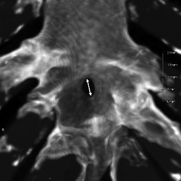
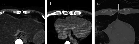
Results: Sternal foramina were present in 3.7% of adults. They were more frequent in males (4.3%) than in females (2.5%), although the difference was insignificant. Most foramina were located at the level of the fifth costal notch and overlaid the pericardium or lung in 72% of evaluable cases. The median diameter was 4.5 mm. In pediatric cases, 11 (12%) exhibited lower-sternebral ossification-center patterns that could form sternal foramina, supporting a developmental origin. An estimated 31% of these patterns may persist into adulthood with unfused segments.
Conclusion: Sternal foramina occurred in 3.7% of adults and were often situated over vital structures, posing procedural risks. Among pediatrics, ossification patterns that may impede fusion-defined as horizontal two-center or ≥ 3 center configurations-were present in 12%, and approximately 31% of these patterns appear to persist into adulthood as sternal foramina. These findings support a developmental basis for sternal foramina and emphasize the importance of recognizing them during imaging and procedural planning.
期刊介绍:
Radiology Research and Practice is a peer-reviewed, Open Access journal that publishes articles on all areas of medical imaging. The journal promotes evidence-based radiology practice though the publication of original research, reviews, and clinical studies for a multidisciplinary audience. Radiology Research and Practice is archived in Portico, which provides permanent archiving for electronic scholarly journals, as well as via the LOCKSS initiative. It operates a fully open access publishing model which allows open global access to its published content. This model is supported through Article Processing Charges. For more information on Article Processing charges in gen


 求助内容:
求助内容: 应助结果提醒方式:


