J Lee Pace, Tanner E James, Nicholas R Anable, Olubusola A Brimmo, John P Abt
{"title":"滑车外侧2位图像倾斜判断滑车发育不良所致髌骨不稳定的有效性。","authors":"J Lee Pace, Tanner E James, Nicholas R Anable, Olubusola A Brimmo, John P Abt","doi":"10.1177/23259671251371276","DOIUrl":null,"url":null,"abstract":"<p><strong>Background: </strong>Trochlear dysplasia is frequently seen in young patients and can be a cause of patellar instability (PI). A lateral trochlear inclination (LTI) of 11° or less has long been utilized to discriminate between patients with and without some degree of trochlear dysplasia that is likely to contribute to recurrent PI. The traditional methodology for this radiographic measure has been described via a single-image technique. Recently, a 2-image technique was introduced to better account for distal femoral orientation relative to proximal trochlear cartilage. However, further work is required to determine the validity of the 2-image LTI technique in introducing a new diagnostic threshold value for trochlear dysplasia.</p><p><strong>Purpose/hypothesis: </strong>The purpose of this study was to evaluate the validity of the novel 2-image LTI measurement technique in determining a diagnostic cut-off value for trochlear dysplasia that is likely to result in recurrent PI. It was hypothesized that this would yield a different value than the historical 11° cutoff determined through the single-image technique.</p><p><strong>Study design: </strong>Cohort study (Diagnosis); Level of evidence, 3.</p><p><strong>Methods: </strong>After institutional review board approval, medical records were retrospectively reviewed to form a PI group and a control (no PI) group. The 2-image LTI was first measured by 2 raters to determine intrarater and interrater reliability. A receiver operating characteristic curve was created to determine a diagnostic cut-off value. After applying this cutoff between the groups, the resultant sensitivity and specificity were calculated.</p><p><strong>Results: </strong>The 2-image LTI measurements demonstrated excellent intrarater and interrater reliability. The PI group had a mean LTI of 2.58°, while the control group had a mean LTI of 17.26°. A diagnostic threshold LTI value of 13.7° was determined, discriminating between patients with and without recurrent PI due to trochlear dysplasia, with a sensitivity of 0.889 and a specificity of 0.780. The area under the curve was 0.887.</p><p><strong>Conclusion: </strong>Utilizing the 2-image LTI measurement technique, a threshold value of 13.7° was determined as optimal for discerning between patients with and without trochlear dysplasia that is likely to lead to recurrent PI. This value can thus be used for accurate diagnoses of trochlear dysplasia, which can help to inform management, patient counseling, and research in the future.</p>","PeriodicalId":19646,"journal":{"name":"Orthopaedic Journal of Sports Medicine","volume":"13 10","pages":"23259671251371276"},"PeriodicalIF":2.5000,"publicationDate":"2025-10-06","publicationTypes":"Journal Article","fieldsOfStudy":null,"isOpenAccess":false,"openAccessPdf":"https://www.ncbi.nlm.nih.gov/pmc/articles/PMC12501464/pdf/","citationCount":"0","resultStr":"{\"title\":\"Validity of the 2-Image Lateral Trochlear Inclination to Determine Patellar Instability Due to Trochlear Dysplasia.\",\"authors\":\"J Lee Pace, Tanner E James, Nicholas R Anable, Olubusola A Brimmo, John P Abt\",\"doi\":\"10.1177/23259671251371276\",\"DOIUrl\":null,\"url\":null,\"abstract\":\"<p><strong>Background: </strong>Trochlear dysplasia is frequently seen in young patients and can be a cause of patellar instability (PI). A lateral trochlear inclination (LTI) of 11° or less has long been utilized to discriminate between patients with and without some degree of trochlear dysplasia that is likely to contribute to recurrent PI. The traditional methodology for this radiographic measure has been described via a single-image technique. Recently, a 2-image technique was introduced to better account for distal femoral orientation relative to proximal trochlear cartilage. However, further work is required to determine the validity of the 2-image LTI technique in introducing a new diagnostic threshold value for trochlear dysplasia.</p><p><strong>Purpose/hypothesis: </strong>The purpose of this study was to evaluate the validity of the novel 2-image LTI measurement technique in determining a diagnostic cut-off value for trochlear dysplasia that is likely to result in recurrent PI. It was hypothesized that this would yield a different value than the historical 11° cutoff determined through the single-image technique.</p><p><strong>Study design: </strong>Cohort study (Diagnosis); Level of evidence, 3.</p><p><strong>Methods: </strong>After institutional review board approval, medical records were retrospectively reviewed to form a PI group and a control (no PI) group. The 2-image LTI was first measured by 2 raters to determine intrarater and interrater reliability. A receiver operating characteristic curve was created to determine a diagnostic cut-off value. After applying this cutoff between the groups, the resultant sensitivity and specificity were calculated.</p><p><strong>Results: </strong>The 2-image LTI measurements demonstrated excellent intrarater and interrater reliability. The PI group had a mean LTI of 2.58°, while the control group had a mean LTI of 17.26°. A diagnostic threshold LTI value of 13.7° was determined, discriminating between patients with and without recurrent PI due to trochlear dysplasia, with a sensitivity of 0.889 and a specificity of 0.780. The area under the curve was 0.887.</p><p><strong>Conclusion: </strong>Utilizing the 2-image LTI measurement technique, a threshold value of 13.7° was determined as optimal for discerning between patients with and without trochlear dysplasia that is likely to lead to recurrent PI. This value can thus be used for accurate diagnoses of trochlear dysplasia, which can help to inform management, patient counseling, and research in the future.</p>\",\"PeriodicalId\":19646,\"journal\":{\"name\":\"Orthopaedic Journal of Sports Medicine\",\"volume\":\"13 10\",\"pages\":\"23259671251371276\"},\"PeriodicalIF\":2.5000,\"publicationDate\":\"2025-10-06\",\"publicationTypes\":\"Journal Article\",\"fieldsOfStudy\":null,\"isOpenAccess\":false,\"openAccessPdf\":\"https://www.ncbi.nlm.nih.gov/pmc/articles/PMC12501464/pdf/\",\"citationCount\":\"0\",\"resultStr\":null,\"platform\":\"Semanticscholar\",\"paperid\":null,\"PeriodicalName\":\"Orthopaedic Journal of Sports Medicine\",\"FirstCategoryId\":\"3\",\"ListUrlMain\":\"https://doi.org/10.1177/23259671251371276\",\"RegionNum\":3,\"RegionCategory\":\"医学\",\"ArticlePicture\":[],\"TitleCN\":null,\"AbstractTextCN\":null,\"PMCID\":null,\"EPubDate\":\"2025/10/1 0:00:00\",\"PubModel\":\"eCollection\",\"JCR\":\"Q2\",\"JCRName\":\"ORTHOPEDICS\",\"Score\":null,\"Total\":0}","platform":"Semanticscholar","paperid":null,"PeriodicalName":"Orthopaedic Journal of Sports Medicine","FirstCategoryId":"3","ListUrlMain":"https://doi.org/10.1177/23259671251371276","RegionNum":3,"RegionCategory":"医学","ArticlePicture":[],"TitleCN":null,"AbstractTextCN":null,"PMCID":null,"EPubDate":"2025/10/1 0:00:00","PubModel":"eCollection","JCR":"Q2","JCRName":"ORTHOPEDICS","Score":null,"Total":0}
Validity of the 2-Image Lateral Trochlear Inclination to Determine Patellar Instability Due to Trochlear Dysplasia.
Background: Trochlear dysplasia is frequently seen in young patients and can be a cause of patellar instability (PI). A lateral trochlear inclination (LTI) of 11° or less has long been utilized to discriminate between patients with and without some degree of trochlear dysplasia that is likely to contribute to recurrent PI. The traditional methodology for this radiographic measure has been described via a single-image technique. Recently, a 2-image technique was introduced to better account for distal femoral orientation relative to proximal trochlear cartilage. However, further work is required to determine the validity of the 2-image LTI technique in introducing a new diagnostic threshold value for trochlear dysplasia.
Purpose/hypothesis: The purpose of this study was to evaluate the validity of the novel 2-image LTI measurement technique in determining a diagnostic cut-off value for trochlear dysplasia that is likely to result in recurrent PI. It was hypothesized that this would yield a different value than the historical 11° cutoff determined through the single-image technique.
Study design: Cohort study (Diagnosis); Level of evidence, 3.
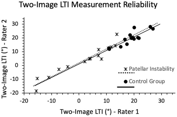
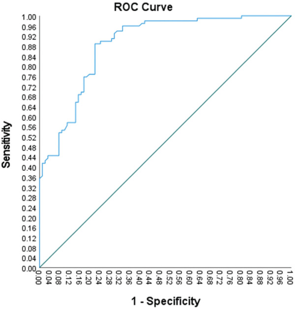
Methods: After institutional review board approval, medical records were retrospectively reviewed to form a PI group and a control (no PI) group. The 2-image LTI was first measured by 2 raters to determine intrarater and interrater reliability. A receiver operating characteristic curve was created to determine a diagnostic cut-off value. After applying this cutoff between the groups, the resultant sensitivity and specificity were calculated.
Results: The 2-image LTI measurements demonstrated excellent intrarater and interrater reliability. The PI group had a mean LTI of 2.58°, while the control group had a mean LTI of 17.26°. A diagnostic threshold LTI value of 13.7° was determined, discriminating between patients with and without recurrent PI due to trochlear dysplasia, with a sensitivity of 0.889 and a specificity of 0.780. The area under the curve was 0.887.
Conclusion: Utilizing the 2-image LTI measurement technique, a threshold value of 13.7° was determined as optimal for discerning between patients with and without trochlear dysplasia that is likely to lead to recurrent PI. This value can thus be used for accurate diagnoses of trochlear dysplasia, which can help to inform management, patient counseling, and research in the future.
期刊介绍:
The Orthopaedic Journal of Sports Medicine (OJSM), developed by the American Orthopaedic Society for Sports Medicine (AOSSM), is a global, peer-reviewed, open access journal that combines the interests of researchers and clinical practitioners across orthopaedic sports medicine, arthroscopy, and knee arthroplasty.
Topics include original research in the areas of:
-Orthopaedic Sports Medicine, including surgical and nonsurgical treatment of orthopaedic sports injuries
-Arthroscopic Surgery (Shoulder/Elbow/Wrist/Hip/Knee/Ankle/Foot)
-Relevant translational research
-Sports traumatology/epidemiology
-Knee and shoulder arthroplasty
The OJSM also publishes relevant systematic reviews and meta-analyses.
This journal is a member of the Committee on Publication Ethics (COPE).

 求助内容:
求助内容: 应助结果提醒方式:


