{"title":"溃疡性结肠炎患者对整合素抑制剂治疗反应的组织学预测因素。","authors":"Takuya Kimizuka, Atsushi Yoshida, Fumiaki Ueno, Yutaka Endo, Yo Kato, Katsuyoshi Matsuoka, Tadakazu Hisamatsu, Toshifumi Hibi","doi":"10.1159/000547013","DOIUrl":null,"url":null,"abstract":"<p><strong>Introduction: </strong>It is crucial to predict the effectiveness of advanced therapies before their administration in ulcerative colitis (UC). Only a few studies have revealed predictive histological factors. Here, we sought to determine whether conventional histology of pretreatment endoscopic biopsy specimens can predict the response to integrin inhibitors.</p><p><strong>Methods: </strong>In the present single-center retrospective study, we examined histopathological findings before initiating an integrin inhibitor. Primary response (PR) was defined as a ≥3-point decrease in the partial Mayo score at 14 weeks. Logistic regression was used to identify the factors predictive for a PR.</p><p><strong>Results: </strong>We analyzed 21 biological and Janus kinase inhibitor-naïve patients with UC. The median baseline Mayo score was 7 (IQR, 6-8), and the C-reactive protein was 0.36 (IQR, 0.11-0.74). Histological findings included large lymphoid follicles (LF) in 61.9% (13/21), basal plasma cell infiltration in 52.4% (11/21), and eosinophilic infiltration (EO) in 42.9% (9/21). PR at 14 weeks was achieved in 57.1% (12/21). Among PR patients, LF was present in 91.7% (11/12), BP in 41.7% (5/12), and EO in 25.0% (3/12). PR was observed in 76.9% (10/13) of LF-positive patients vs. 12.5% (1/8) of LF-negative patients (<i>p</i> = 0.01). LF was significantly associated with the response to integrin inhibitors, whereas BP and EO were not.</p><p><strong>Conclusion: </strong>The presence of LF in biopsy specimens predicts the response to integrin inhibitors in patients with UC. Conventional histological examinations may aid in predicting therapeutic responses to advanced therapies.</p>","PeriodicalId":13605,"journal":{"name":"Inflammatory Intestinal Diseases","volume":"10 1","pages":"224-232"},"PeriodicalIF":0.0000,"publicationDate":"2025-08-28","publicationTypes":"Journal Article","fieldsOfStudy":null,"isOpenAccess":false,"openAccessPdf":"https://www.ncbi.nlm.nih.gov/pmc/articles/PMC12503538/pdf/","citationCount":"0","resultStr":"{\"title\":\"Histological Predictors for Therapeutic Response to Integrin Inhibitors in Patients with Ulcerative Colitis.\",\"authors\":\"Takuya Kimizuka, Atsushi Yoshida, Fumiaki Ueno, Yutaka Endo, Yo Kato, Katsuyoshi Matsuoka, Tadakazu Hisamatsu, Toshifumi Hibi\",\"doi\":\"10.1159/000547013\",\"DOIUrl\":null,\"url\":null,\"abstract\":\"<p><strong>Introduction: </strong>It is crucial to predict the effectiveness of advanced therapies before their administration in ulcerative colitis (UC). Only a few studies have revealed predictive histological factors. Here, we sought to determine whether conventional histology of pretreatment endoscopic biopsy specimens can predict the response to integrin inhibitors.</p><p><strong>Methods: </strong>In the present single-center retrospective study, we examined histopathological findings before initiating an integrin inhibitor. Primary response (PR) was defined as a ≥3-point decrease in the partial Mayo score at 14 weeks. Logistic regression was used to identify the factors predictive for a PR.</p><p><strong>Results: </strong>We analyzed 21 biological and Janus kinase inhibitor-naïve patients with UC. The median baseline Mayo score was 7 (IQR, 6-8), and the C-reactive protein was 0.36 (IQR, 0.11-0.74). Histological findings included large lymphoid follicles (LF) in 61.9% (13/21), basal plasma cell infiltration in 52.4% (11/21), and eosinophilic infiltration (EO) in 42.9% (9/21). PR at 14 weeks was achieved in 57.1% (12/21). Among PR patients, LF was present in 91.7% (11/12), BP in 41.7% (5/12), and EO in 25.0% (3/12). PR was observed in 76.9% (10/13) of LF-positive patients vs. 12.5% (1/8) of LF-negative patients (<i>p</i> = 0.01). LF was significantly associated with the response to integrin inhibitors, whereas BP and EO were not.</p><p><strong>Conclusion: </strong>The presence of LF in biopsy specimens predicts the response to integrin inhibitors in patients with UC. Conventional histological examinations may aid in predicting therapeutic responses to advanced therapies.</p>\",\"PeriodicalId\":13605,\"journal\":{\"name\":\"Inflammatory Intestinal Diseases\",\"volume\":\"10 1\",\"pages\":\"224-232\"},\"PeriodicalIF\":0.0000,\"publicationDate\":\"2025-08-28\",\"publicationTypes\":\"Journal Article\",\"fieldsOfStudy\":null,\"isOpenAccess\":false,\"openAccessPdf\":\"https://www.ncbi.nlm.nih.gov/pmc/articles/PMC12503538/pdf/\",\"citationCount\":\"0\",\"resultStr\":null,\"platform\":\"Semanticscholar\",\"paperid\":null,\"PeriodicalName\":\"Inflammatory Intestinal Diseases\",\"FirstCategoryId\":\"1085\",\"ListUrlMain\":\"https://doi.org/10.1159/000547013\",\"RegionNum\":0,\"RegionCategory\":null,\"ArticlePicture\":[],\"TitleCN\":null,\"AbstractTextCN\":null,\"PMCID\":null,\"EPubDate\":\"2025/1/1 0:00:00\",\"PubModel\":\"eCollection\",\"JCR\":\"Q2\",\"JCRName\":\"Medicine\",\"Score\":null,\"Total\":0}","platform":"Semanticscholar","paperid":null,"PeriodicalName":"Inflammatory Intestinal Diseases","FirstCategoryId":"1085","ListUrlMain":"https://doi.org/10.1159/000547013","RegionNum":0,"RegionCategory":null,"ArticlePicture":[],"TitleCN":null,"AbstractTextCN":null,"PMCID":null,"EPubDate":"2025/1/1 0:00:00","PubModel":"eCollection","JCR":"Q2","JCRName":"Medicine","Score":null,"Total":0}
Histological Predictors for Therapeutic Response to Integrin Inhibitors in Patients with Ulcerative Colitis.
Introduction: It is crucial to predict the effectiveness of advanced therapies before their administration in ulcerative colitis (UC). Only a few studies have revealed predictive histological factors. Here, we sought to determine whether conventional histology of pretreatment endoscopic biopsy specimens can predict the response to integrin inhibitors.
Methods: In the present single-center retrospective study, we examined histopathological findings before initiating an integrin inhibitor. Primary response (PR) was defined as a ≥3-point decrease in the partial Mayo score at 14 weeks. Logistic regression was used to identify the factors predictive for a PR.
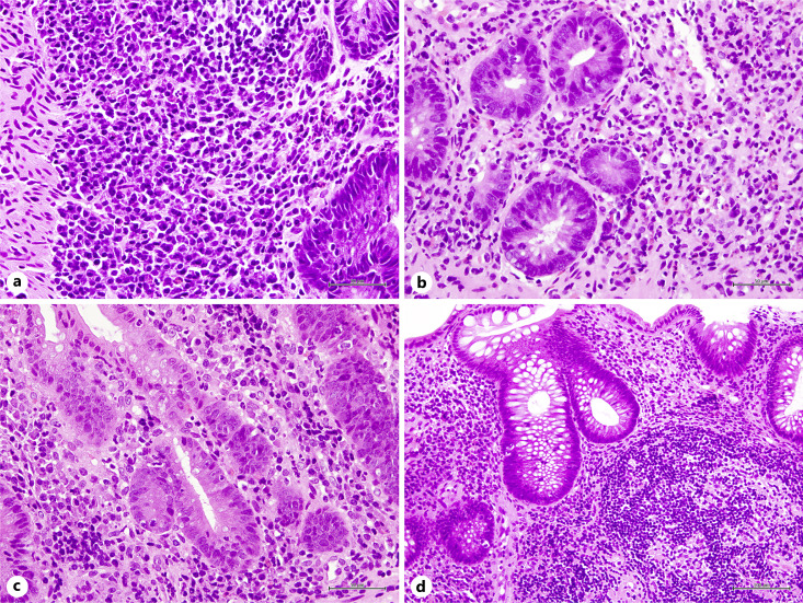
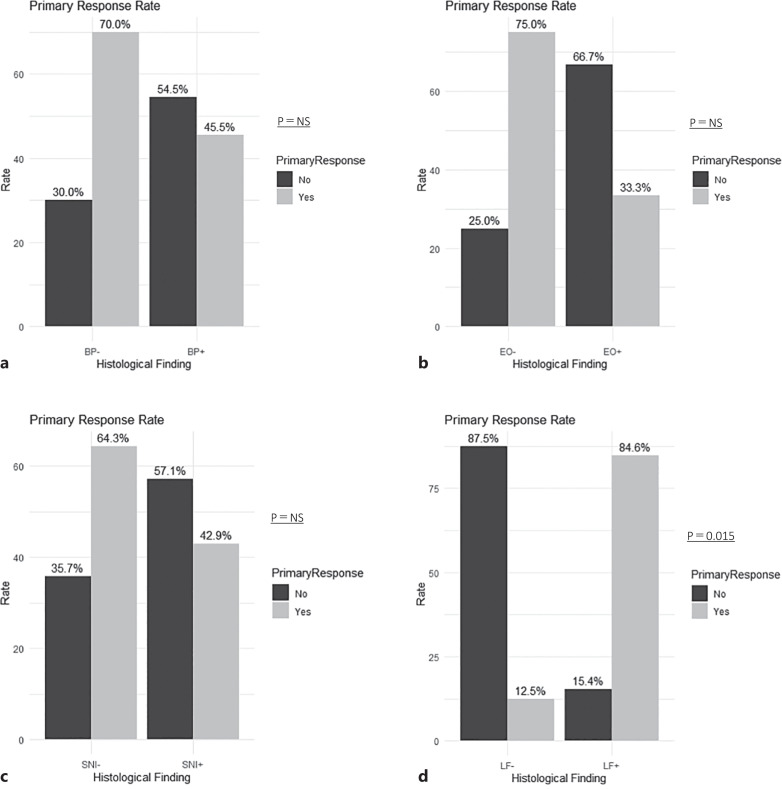
Results: We analyzed 21 biological and Janus kinase inhibitor-naïve patients with UC. The median baseline Mayo score was 7 (IQR, 6-8), and the C-reactive protein was 0.36 (IQR, 0.11-0.74). Histological findings included large lymphoid follicles (LF) in 61.9% (13/21), basal plasma cell infiltration in 52.4% (11/21), and eosinophilic infiltration (EO) in 42.9% (9/21). PR at 14 weeks was achieved in 57.1% (12/21). Among PR patients, LF was present in 91.7% (11/12), BP in 41.7% (5/12), and EO in 25.0% (3/12). PR was observed in 76.9% (10/13) of LF-positive patients vs. 12.5% (1/8) of LF-negative patients (p = 0.01). LF was significantly associated with the response to integrin inhibitors, whereas BP and EO were not.
Conclusion: The presence of LF in biopsy specimens predicts the response to integrin inhibitors in patients with UC. Conventional histological examinations may aid in predicting therapeutic responses to advanced therapies.

 求助内容:
求助内容: 应助结果提醒方式:


