{"title":"使用低剂量CT灌注量化下肢血流:猪模型验证。","authors":"Alireza Shojazadeh, Negin Hadjiabdolhamid, Dale J Black, Ines Antunes, Chaeeun Lee, Wenbo Li, Sabee Molloi","doi":"10.1093/radadv/umae029","DOIUrl":null,"url":null,"abstract":"<p><strong>Background: </strong>Quantitative assessment of blood flow in peripheral extremities in conjunction with simultaneous CT angiography measurements can improve risk assessment and provide a critical decision-making tool for patients across a wide spectrum of vascular disease severity.</p><p><strong>Purpose: </strong>This study assessed the reproducibility and accuracy of lower extremity blood flow measurements with a low-dose first-pass analysis CT perfusion technique.</p><p><strong>Materials and methods: </strong>This prospective study utilized 16 Yorkshire Swine to obtain lower extremity blood flow CT measurements at baseline and under induced femoral stenosis using a vascular occluder. Thirty-three pairs of CT measurements evaluated reproducibility, and 43 CT measurements assessed accuracy against ultrasound flow probe references. Contrast agent and saline chaser were both injected peripherally at a rate of 5 mL/s. Bolus tracking was used, and a pre-contrast and post-contrast helical scan were acquired at the base and approximately the peak of the femoral enhancement (CT angiogram), respectively. The acquired data were then used as analytical inputs into a first-pass analysis model to derive perfusion in mL/min/g. The reproducibility and accuracy of lower extremity perfusion measurements were assessed via Mixed model regression and Bland-Altman analysis.</p><p><strong>Results: </strong>Calculated CT perfusion measurements derived from first-pass analysis technique (<i>P</i> <sub>CT</sub>), and the reference standard ultrasound perfusion measurements (<i>P</i> <sub>ref</sub>) were related by <i>P</i> <sub>CT</sub> = 1.06 <i>P</i> <sub>ref</sub> + 0.00 (<i>r</i> <sup>2</sup> = 0.90, Root-Mean-Square Error [RMSE] = 0.01 mL/min/g). The first (<i>P</i> <sub>1</sub>) and second (<i>P</i> <sub>2</sub>) CT perfusion measurements were related by <i>P</i> <sub>2</sub> = 0.98 <i>P</i> <sub>1</sub> + 0.02 (<i>r</i> = 0.97, RMSE = 0.11 mL/min/g). The average effective dose of perfusion measurement using first-pass analysis technique was calculated to be only 2.13 mSv.</p><p><strong>Conclusion: </strong>The low-dose quantitative CT perfusion technique can accurately measure lower extremity perfusion (mL/min/g) using only 2 helical scans. The CT angiogram and perfusion measurements can be used as a comprehensive technique for morphological and physiological assessment of limb ischemia.</p>","PeriodicalId":519940,"journal":{"name":"Radiology advances","volume":"1 4","pages":"umae029"},"PeriodicalIF":0.0000,"publicationDate":"2024-11-09","publicationTypes":"Journal Article","fieldsOfStudy":null,"isOpenAccess":false,"openAccessPdf":"https://www.ncbi.nlm.nih.gov/pmc/articles/PMC12429254/pdf/","citationCount":"0","resultStr":"{\"title\":\"Quantifying lower extremity blood flow using low-dose CT perfusion: validation in a swine model.\",\"authors\":\"Alireza Shojazadeh, Negin Hadjiabdolhamid, Dale J Black, Ines Antunes, Chaeeun Lee, Wenbo Li, Sabee Molloi\",\"doi\":\"10.1093/radadv/umae029\",\"DOIUrl\":null,\"url\":null,\"abstract\":\"<p><strong>Background: </strong>Quantitative assessment of blood flow in peripheral extremities in conjunction with simultaneous CT angiography measurements can improve risk assessment and provide a critical decision-making tool for patients across a wide spectrum of vascular disease severity.</p><p><strong>Purpose: </strong>This study assessed the reproducibility and accuracy of lower extremity blood flow measurements with a low-dose first-pass analysis CT perfusion technique.</p><p><strong>Materials and methods: </strong>This prospective study utilized 16 Yorkshire Swine to obtain lower extremity blood flow CT measurements at baseline and under induced femoral stenosis using a vascular occluder. Thirty-three pairs of CT measurements evaluated reproducibility, and 43 CT measurements assessed accuracy against ultrasound flow probe references. Contrast agent and saline chaser were both injected peripherally at a rate of 5 mL/s. Bolus tracking was used, and a pre-contrast and post-contrast helical scan were acquired at the base and approximately the peak of the femoral enhancement (CT angiogram), respectively. The acquired data were then used as analytical inputs into a first-pass analysis model to derive perfusion in mL/min/g. The reproducibility and accuracy of lower extremity perfusion measurements were assessed via Mixed model regression and Bland-Altman analysis.</p><p><strong>Results: </strong>Calculated CT perfusion measurements derived from first-pass analysis technique (<i>P</i> <sub>CT</sub>), and the reference standard ultrasound perfusion measurements (<i>P</i> <sub>ref</sub>) were related by <i>P</i> <sub>CT</sub> = 1.06 <i>P</i> <sub>ref</sub> + 0.00 (<i>r</i> <sup>2</sup> = 0.90, Root-Mean-Square Error [RMSE] = 0.01 mL/min/g). The first (<i>P</i> <sub>1</sub>) and second (<i>P</i> <sub>2</sub>) CT perfusion measurements were related by <i>P</i> <sub>2</sub> = 0.98 <i>P</i> <sub>1</sub> + 0.02 (<i>r</i> = 0.97, RMSE = 0.11 mL/min/g). The average effective dose of perfusion measurement using first-pass analysis technique was calculated to be only 2.13 mSv.</p><p><strong>Conclusion: </strong>The low-dose quantitative CT perfusion technique can accurately measure lower extremity perfusion (mL/min/g) using only 2 helical scans. The CT angiogram and perfusion measurements can be used as a comprehensive technique for morphological and physiological assessment of limb ischemia.</p>\",\"PeriodicalId\":519940,\"journal\":{\"name\":\"Radiology advances\",\"volume\":\"1 4\",\"pages\":\"umae029\"},\"PeriodicalIF\":0.0000,\"publicationDate\":\"2024-11-09\",\"publicationTypes\":\"Journal Article\",\"fieldsOfStudy\":null,\"isOpenAccess\":false,\"openAccessPdf\":\"https://www.ncbi.nlm.nih.gov/pmc/articles/PMC12429254/pdf/\",\"citationCount\":\"0\",\"resultStr\":null,\"platform\":\"Semanticscholar\",\"paperid\":null,\"PeriodicalName\":\"Radiology advances\",\"FirstCategoryId\":\"1085\",\"ListUrlMain\":\"https://doi.org/10.1093/radadv/umae029\",\"RegionNum\":0,\"RegionCategory\":null,\"ArticlePicture\":[],\"TitleCN\":null,\"AbstractTextCN\":null,\"PMCID\":null,\"EPubDate\":\"2024/11/1 0:00:00\",\"PubModel\":\"eCollection\",\"JCR\":\"\",\"JCRName\":\"\",\"Score\":null,\"Total\":0}","platform":"Semanticscholar","paperid":null,"PeriodicalName":"Radiology advances","FirstCategoryId":"1085","ListUrlMain":"https://doi.org/10.1093/radadv/umae029","RegionNum":0,"RegionCategory":null,"ArticlePicture":[],"TitleCN":null,"AbstractTextCN":null,"PMCID":null,"EPubDate":"2024/11/1 0:00:00","PubModel":"eCollection","JCR":"","JCRName":"","Score":null,"Total":0}
Quantifying lower extremity blood flow using low-dose CT perfusion: validation in a swine model.
Background: Quantitative assessment of blood flow in peripheral extremities in conjunction with simultaneous CT angiography measurements can improve risk assessment and provide a critical decision-making tool for patients across a wide spectrum of vascular disease severity.
Purpose: This study assessed the reproducibility and accuracy of lower extremity blood flow measurements with a low-dose first-pass analysis CT perfusion technique.
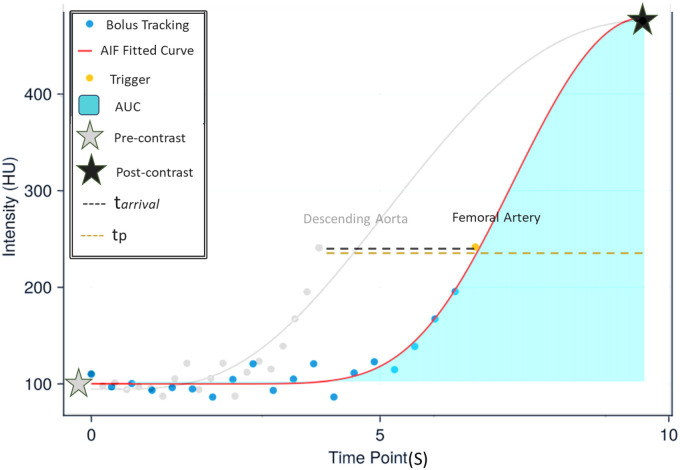
Materials and methods: This prospective study utilized 16 Yorkshire Swine to obtain lower extremity blood flow CT measurements at baseline and under induced femoral stenosis using a vascular occluder. Thirty-three pairs of CT measurements evaluated reproducibility, and 43 CT measurements assessed accuracy against ultrasound flow probe references. Contrast agent and saline chaser were both injected peripherally at a rate of 5 mL/s. Bolus tracking was used, and a pre-contrast and post-contrast helical scan were acquired at the base and approximately the peak of the femoral enhancement (CT angiogram), respectively. The acquired data were then used as analytical inputs into a first-pass analysis model to derive perfusion in mL/min/g. The reproducibility and accuracy of lower extremity perfusion measurements were assessed via Mixed model regression and Bland-Altman analysis.
Results: Calculated CT perfusion measurements derived from first-pass analysis technique (PCT), and the reference standard ultrasound perfusion measurements (Pref) were related by PCT = 1.06 Pref + 0.00 (r2 = 0.90, Root-Mean-Square Error [RMSE] = 0.01 mL/min/g). The first (P1) and second (P2) CT perfusion measurements were related by P2 = 0.98 P1 + 0.02 (r = 0.97, RMSE = 0.11 mL/min/g). The average effective dose of perfusion measurement using first-pass analysis technique was calculated to be only 2.13 mSv.
Conclusion: The low-dose quantitative CT perfusion technique can accurately measure lower extremity perfusion (mL/min/g) using only 2 helical scans. The CT angiogram and perfusion measurements can be used as a comprehensive technique for morphological and physiological assessment of limb ischemia.

 求助内容:
求助内容: 应助结果提醒方式:


