Raj Kumar Panta, Zhye Yin, Fredrik Grönberg, Benjamin Wildman-Tobriner, Mridul Bhattarai, Ehsan Abadi, Paul Segars, Ehsan Samei
{"title":"用深硅光子计数CT定量肝脏脂肪:一项硅成像研究。","authors":"Raj Kumar Panta, Zhye Yin, Fredrik Grönberg, Benjamin Wildman-Tobriner, Mridul Bhattarai, Ehsan Abadi, Paul Segars, Ehsan Samei","doi":"10.1093/radadv/umaf031","DOIUrl":null,"url":null,"abstract":"<p><strong>Background: </strong>Accurate liver fat quantification is essential for early diagnosis and effective management of fatty liver disease.</p><p><strong>Purpose: </strong>To investigate the potential clinical utility of a deep silicon-based photon-counting CT (dSi-PCCT), currently in development, for liver fat quantification using human models in an <i>in silico</i> imaging study.</p><p><strong>Materials and methods: </strong>dSi-PCCT is a cutting-edge photon-counting CT (GE HealthCare), with several investigational systems installed globally, used under IRB approval for imaging animals and human volunteers to support FDA clearance. We developed a dSi-PCCT simulator and benchmarked its imaging performance with respect to a prototype. We imaged a computational Gammex phantom with fat fractions (FF) ranging from 0% to 100%, along with five XCAT human models with liver FF ranging from 1% to 50%, using an abdominal CT protocol. The resulting spectral sinograms were processed using a material decomposition (MD) technique. We calculated HU-based Proton Density Fat Fraction (PDFF) from single-energy images in XCAT models and compared it against the MD-derived FF. The MD-derived FF of both datasets was assessed against the digitally defined ground truth values.</p><p><strong>Results: </strong>We observed a strong correlation (<i>R</i> <sup>2</sup> = 0.98) between MD-derived, HU-based PDFF, and ground-truth FF in a Gammex and XCAT models. There was no statistically significant difference (<i>P</i> = .52) in FF quantification accuracy between Gammex and the XCAT human models. The root mean square errors were 4.7% for Gammex and 2.7% for XCAT. Bland-Altman analysis further confirmed good agreement between the ground truth and MD-derived FF, with differences in FF ranging from -6.9% to 7% for Gammex and -3.0% to 37.6% for XCAT.</p><p><strong>Conclusion: </strong>The results indicate that dSi-PCCT could enable accurate liver fat quantification across a wide range of FFs in multiple objects. These findings suggest that the potential utility of dSi-PCCT for accurate liver fat assessment should be explored <i>in vivo</i>.</p>","PeriodicalId":519940,"journal":{"name":"Radiology advances","volume":"2 5","pages":"umaf031"},"PeriodicalIF":0.0000,"publicationDate":"2025-09-02","publicationTypes":"Journal Article","fieldsOfStudy":null,"isOpenAccess":false,"openAccessPdf":"https://www.ncbi.nlm.nih.gov/pmc/articles/PMC12483155/pdf/","citationCount":"0","resultStr":"{\"title\":\"Liver fat quantification using deep silicon photon-counting CT: an <i>in silico</i> imaging study.\",\"authors\":\"Raj Kumar Panta, Zhye Yin, Fredrik Grönberg, Benjamin Wildman-Tobriner, Mridul Bhattarai, Ehsan Abadi, Paul Segars, Ehsan Samei\",\"doi\":\"10.1093/radadv/umaf031\",\"DOIUrl\":null,\"url\":null,\"abstract\":\"<p><strong>Background: </strong>Accurate liver fat quantification is essential for early diagnosis and effective management of fatty liver disease.</p><p><strong>Purpose: </strong>To investigate the potential clinical utility of a deep silicon-based photon-counting CT (dSi-PCCT), currently in development, for liver fat quantification using human models in an <i>in silico</i> imaging study.</p><p><strong>Materials and methods: </strong>dSi-PCCT is a cutting-edge photon-counting CT (GE HealthCare), with several investigational systems installed globally, used under IRB approval for imaging animals and human volunteers to support FDA clearance. We developed a dSi-PCCT simulator and benchmarked its imaging performance with respect to a prototype. We imaged a computational Gammex phantom with fat fractions (FF) ranging from 0% to 100%, along with five XCAT human models with liver FF ranging from 1% to 50%, using an abdominal CT protocol. The resulting spectral sinograms were processed using a material decomposition (MD) technique. We calculated HU-based Proton Density Fat Fraction (PDFF) from single-energy images in XCAT models and compared it against the MD-derived FF. The MD-derived FF of both datasets was assessed against the digitally defined ground truth values.</p><p><strong>Results: </strong>We observed a strong correlation (<i>R</i> <sup>2</sup> = 0.98) between MD-derived, HU-based PDFF, and ground-truth FF in a Gammex and XCAT models. There was no statistically significant difference (<i>P</i> = .52) in FF quantification accuracy between Gammex and the XCAT human models. The root mean square errors were 4.7% for Gammex and 2.7% for XCAT. Bland-Altman analysis further confirmed good agreement between the ground truth and MD-derived FF, with differences in FF ranging from -6.9% to 7% for Gammex and -3.0% to 37.6% for XCAT.</p><p><strong>Conclusion: </strong>The results indicate that dSi-PCCT could enable accurate liver fat quantification across a wide range of FFs in multiple objects. These findings suggest that the potential utility of dSi-PCCT for accurate liver fat assessment should be explored <i>in vivo</i>.</p>\",\"PeriodicalId\":519940,\"journal\":{\"name\":\"Radiology advances\",\"volume\":\"2 5\",\"pages\":\"umaf031\"},\"PeriodicalIF\":0.0000,\"publicationDate\":\"2025-09-02\",\"publicationTypes\":\"Journal Article\",\"fieldsOfStudy\":null,\"isOpenAccess\":false,\"openAccessPdf\":\"https://www.ncbi.nlm.nih.gov/pmc/articles/PMC12483155/pdf/\",\"citationCount\":\"0\",\"resultStr\":null,\"platform\":\"Semanticscholar\",\"paperid\":null,\"PeriodicalName\":\"Radiology advances\",\"FirstCategoryId\":\"1085\",\"ListUrlMain\":\"https://doi.org/10.1093/radadv/umaf031\",\"RegionNum\":0,\"RegionCategory\":null,\"ArticlePicture\":[],\"TitleCN\":null,\"AbstractTextCN\":null,\"PMCID\":null,\"EPubDate\":\"2025/9/1 0:00:00\",\"PubModel\":\"eCollection\",\"JCR\":\"\",\"JCRName\":\"\",\"Score\":null,\"Total\":0}","platform":"Semanticscholar","paperid":null,"PeriodicalName":"Radiology advances","FirstCategoryId":"1085","ListUrlMain":"https://doi.org/10.1093/radadv/umaf031","RegionNum":0,"RegionCategory":null,"ArticlePicture":[],"TitleCN":null,"AbstractTextCN":null,"PMCID":null,"EPubDate":"2025/9/1 0:00:00","PubModel":"eCollection","JCR":"","JCRName":"","Score":null,"Total":0}
Liver fat quantification using deep silicon photon-counting CT: an in silico imaging study.
Background: Accurate liver fat quantification is essential for early diagnosis and effective management of fatty liver disease.
Purpose: To investigate the potential clinical utility of a deep silicon-based photon-counting CT (dSi-PCCT), currently in development, for liver fat quantification using human models in an in silico imaging study.
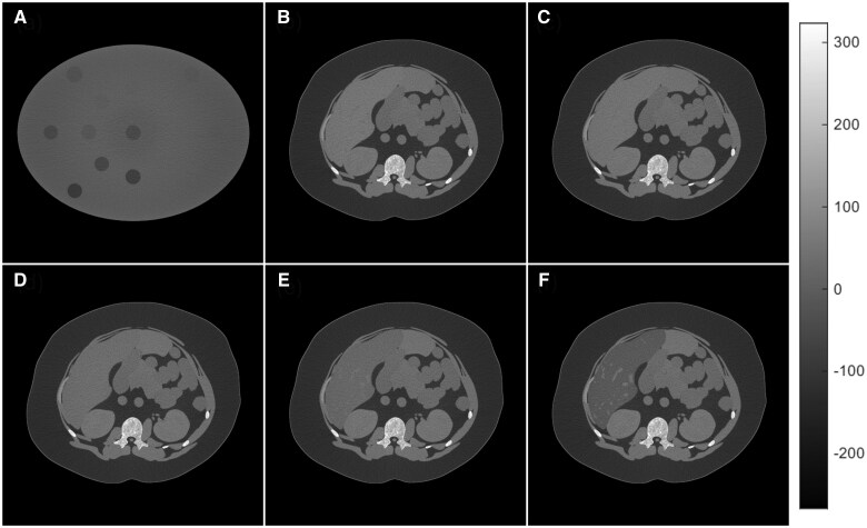
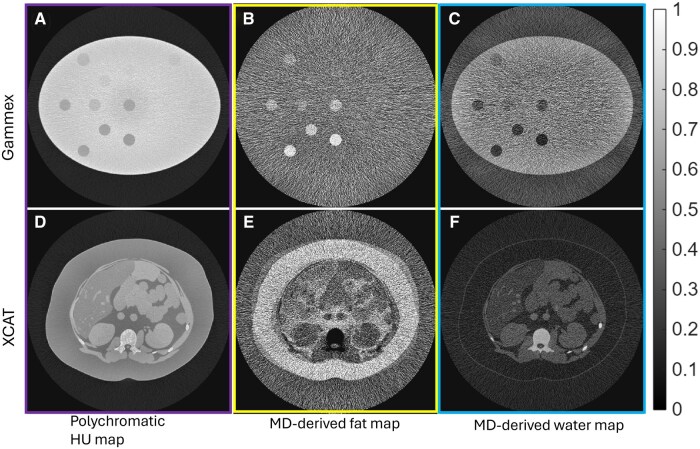
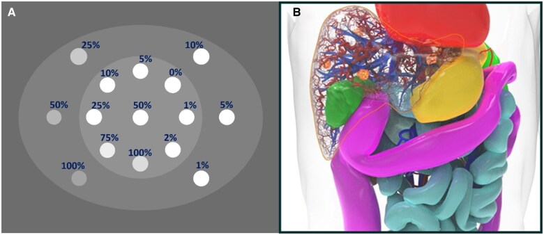
Materials and methods: dSi-PCCT is a cutting-edge photon-counting CT (GE HealthCare), with several investigational systems installed globally, used under IRB approval for imaging animals and human volunteers to support FDA clearance. We developed a dSi-PCCT simulator and benchmarked its imaging performance with respect to a prototype. We imaged a computational Gammex phantom with fat fractions (FF) ranging from 0% to 100%, along with five XCAT human models with liver FF ranging from 1% to 50%, using an abdominal CT protocol. The resulting spectral sinograms were processed using a material decomposition (MD) technique. We calculated HU-based Proton Density Fat Fraction (PDFF) from single-energy images in XCAT models and compared it against the MD-derived FF. The MD-derived FF of both datasets was assessed against the digitally defined ground truth values.
Results: We observed a strong correlation (R2 = 0.98) between MD-derived, HU-based PDFF, and ground-truth FF in a Gammex and XCAT models. There was no statistically significant difference (P = .52) in FF quantification accuracy between Gammex and the XCAT human models. The root mean square errors were 4.7% for Gammex and 2.7% for XCAT. Bland-Altman analysis further confirmed good agreement between the ground truth and MD-derived FF, with differences in FF ranging from -6.9% to 7% for Gammex and -3.0% to 37.6% for XCAT.
Conclusion: The results indicate that dSi-PCCT could enable accurate liver fat quantification across a wide range of FFs in multiple objects. These findings suggest that the potential utility of dSi-PCCT for accurate liver fat assessment should be explored in vivo.

 求助内容:
求助内容: 应助结果提醒方式:


