Brahim Mehadji, Talia Marx, Adrianna Carter, Roger Eric Goldman, Catherine Tram Vu, Emilie Roncali
{"title":"对比增强CT作为肝动脉放射栓塞中肺分流分数评估的无创替代方法。","authors":"Brahim Mehadji, Talia Marx, Adrianna Carter, Roger Eric Goldman, Catherine Tram Vu, Emilie Roncali","doi":"10.1093/radadv/umaf025","DOIUrl":null,"url":null,"abstract":"<p><strong>Background: </strong>Estimation of the lung shunt fraction (LSF) is an integral part of liver radioembolization treatment planning to prevent excessive lung irradiation from arterio-venous shunting in the liver. <sup>99m</sup>Tc macro-aggregated albumin (<sup>99m</sup>Tc-MAA) nuclear imaging is the standard method. Recent literature suggests that <sup>99m</sup>Tc-MAA nuclear imaging may be omitted in selected patient populations.</p><p><strong>Purpose: </strong>This study investigates the potential of contrast-enhanced computed tomography (CECT) as a non-invasive method for estimating LSF as an alternative for <sup>99m</sup>Tc-MAA nuclear imaging.</p><p><strong>Materials and methods: </strong>This single-center retrospective study included 30 consecutive patients who underwent <sup>90</sup>Y radioembolization between January 2015 and December 2024, where both four-phase CECT and <sup>99m</sup>Tc-MAA planar imaging were performed within one month of each other. Hypervascular tumor enhancement was identified on the CECT by subtracting the portal venous phase from the arterial phase and applying an intensity threshold. Additional perfusion characteristics were captured. Statistical analysis assessed the agreement between the CECT-derived volume ratios and the LSF values derived from <sup>99m</sup>Tc-MAA imaging.</p><p><strong>Results: </strong>The cohort consisted of 23 male and 7 female patients with a median age of 66 years (interquartile range: 58-71), diagnosed with hepatocellular carcinoma (<i>n</i> = 24), intrahepatic cholangiocarcinoma (<i>n</i> = 2), pancreatic neuroendocrine tumors (<i>n</i> = 2), metastatic colorectal cancer (<i>n</i> = 1), and lymphocyte carcinoma (<i>n</i> = 1). Regression of the hypervascular-tumor-to-perfused volume ratio on CECT against LSF from <sup>99m</sup>Tc-MAA imaging showed <i>R</i> <sup>2</sup> = 0.95 (<i>P</i> < .001). In contrast, the correlation between tumor volume and LSF was <i>R</i> <sup>2</sup> = 0.38 (<i>P</i> = .001). The root mean square error between the LSF estimated from CECT and that measured using <sup>99m</sup>Tc-MAA planar imaging was 3%.</p><p><strong>Conclusion: </strong>Hypervascular-tumor-to-perfused volume ratio computed from CECT may offer a suitable alternative to <sup>99m</sup>Tc-MAA nuclear imaging for LSF estimation in patients undergoing transarterial radioembolization.</p>","PeriodicalId":519940,"journal":{"name":"Radiology advances","volume":"2 4","pages":"umaf025"},"PeriodicalIF":0.0000,"publicationDate":"2025-08-04","publicationTypes":"Journal Article","fieldsOfStudy":null,"isOpenAccess":false,"openAccessPdf":"https://www.ncbi.nlm.nih.gov/pmc/articles/PMC12429231/pdf/","citationCount":"0","resultStr":"{\"title\":\"Contrast-enhanced CT as a non-invasive alternative for lung shunt fraction estimation in hepatic transarterial radioembolization.\",\"authors\":\"Brahim Mehadji, Talia Marx, Adrianna Carter, Roger Eric Goldman, Catherine Tram Vu, Emilie Roncali\",\"doi\":\"10.1093/radadv/umaf025\",\"DOIUrl\":null,\"url\":null,\"abstract\":\"<p><strong>Background: </strong>Estimation of the lung shunt fraction (LSF) is an integral part of liver radioembolization treatment planning to prevent excessive lung irradiation from arterio-venous shunting in the liver. <sup>99m</sup>Tc macro-aggregated albumin (<sup>99m</sup>Tc-MAA) nuclear imaging is the standard method. Recent literature suggests that <sup>99m</sup>Tc-MAA nuclear imaging may be omitted in selected patient populations.</p><p><strong>Purpose: </strong>This study investigates the potential of contrast-enhanced computed tomography (CECT) as a non-invasive method for estimating LSF as an alternative for <sup>99m</sup>Tc-MAA nuclear imaging.</p><p><strong>Materials and methods: </strong>This single-center retrospective study included 30 consecutive patients who underwent <sup>90</sup>Y radioembolization between January 2015 and December 2024, where both four-phase CECT and <sup>99m</sup>Tc-MAA planar imaging were performed within one month of each other. Hypervascular tumor enhancement was identified on the CECT by subtracting the portal venous phase from the arterial phase and applying an intensity threshold. Additional perfusion characteristics were captured. Statistical analysis assessed the agreement between the CECT-derived volume ratios and the LSF values derived from <sup>99m</sup>Tc-MAA imaging.</p><p><strong>Results: </strong>The cohort consisted of 23 male and 7 female patients with a median age of 66 years (interquartile range: 58-71), diagnosed with hepatocellular carcinoma (<i>n</i> = 24), intrahepatic cholangiocarcinoma (<i>n</i> = 2), pancreatic neuroendocrine tumors (<i>n</i> = 2), metastatic colorectal cancer (<i>n</i> = 1), and lymphocyte carcinoma (<i>n</i> = 1). Regression of the hypervascular-tumor-to-perfused volume ratio on CECT against LSF from <sup>99m</sup>Tc-MAA imaging showed <i>R</i> <sup>2</sup> = 0.95 (<i>P</i> < .001). In contrast, the correlation between tumor volume and LSF was <i>R</i> <sup>2</sup> = 0.38 (<i>P</i> = .001). The root mean square error between the LSF estimated from CECT and that measured using <sup>99m</sup>Tc-MAA planar imaging was 3%.</p><p><strong>Conclusion: </strong>Hypervascular-tumor-to-perfused volume ratio computed from CECT may offer a suitable alternative to <sup>99m</sup>Tc-MAA nuclear imaging for LSF estimation in patients undergoing transarterial radioembolization.</p>\",\"PeriodicalId\":519940,\"journal\":{\"name\":\"Radiology advances\",\"volume\":\"2 4\",\"pages\":\"umaf025\"},\"PeriodicalIF\":0.0000,\"publicationDate\":\"2025-08-04\",\"publicationTypes\":\"Journal Article\",\"fieldsOfStudy\":null,\"isOpenAccess\":false,\"openAccessPdf\":\"https://www.ncbi.nlm.nih.gov/pmc/articles/PMC12429231/pdf/\",\"citationCount\":\"0\",\"resultStr\":null,\"platform\":\"Semanticscholar\",\"paperid\":null,\"PeriodicalName\":\"Radiology advances\",\"FirstCategoryId\":\"1085\",\"ListUrlMain\":\"https://doi.org/10.1093/radadv/umaf025\",\"RegionNum\":0,\"RegionCategory\":null,\"ArticlePicture\":[],\"TitleCN\":null,\"AbstractTextCN\":null,\"PMCID\":null,\"EPubDate\":\"2025/7/1 0:00:00\",\"PubModel\":\"eCollection\",\"JCR\":\"\",\"JCRName\":\"\",\"Score\":null,\"Total\":0}","platform":"Semanticscholar","paperid":null,"PeriodicalName":"Radiology advances","FirstCategoryId":"1085","ListUrlMain":"https://doi.org/10.1093/radadv/umaf025","RegionNum":0,"RegionCategory":null,"ArticlePicture":[],"TitleCN":null,"AbstractTextCN":null,"PMCID":null,"EPubDate":"2025/7/1 0:00:00","PubModel":"eCollection","JCR":"","JCRName":"","Score":null,"Total":0}
Contrast-enhanced CT as a non-invasive alternative for lung shunt fraction estimation in hepatic transarterial radioembolization.
Background: Estimation of the lung shunt fraction (LSF) is an integral part of liver radioembolization treatment planning to prevent excessive lung irradiation from arterio-venous shunting in the liver. 99mTc macro-aggregated albumin (99mTc-MAA) nuclear imaging is the standard method. Recent literature suggests that 99mTc-MAA nuclear imaging may be omitted in selected patient populations.
Purpose: This study investigates the potential of contrast-enhanced computed tomography (CECT) as a non-invasive method for estimating LSF as an alternative for 99mTc-MAA nuclear imaging.
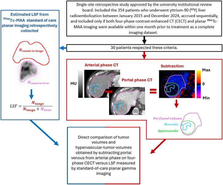
Materials and methods: This single-center retrospective study included 30 consecutive patients who underwent 90Y radioembolization between January 2015 and December 2024, where both four-phase CECT and 99mTc-MAA planar imaging were performed within one month of each other. Hypervascular tumor enhancement was identified on the CECT by subtracting the portal venous phase from the arterial phase and applying an intensity threshold. Additional perfusion characteristics were captured. Statistical analysis assessed the agreement between the CECT-derived volume ratios and the LSF values derived from 99mTc-MAA imaging.
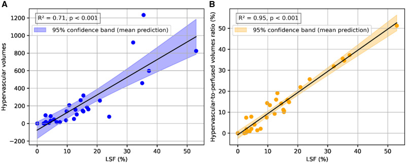
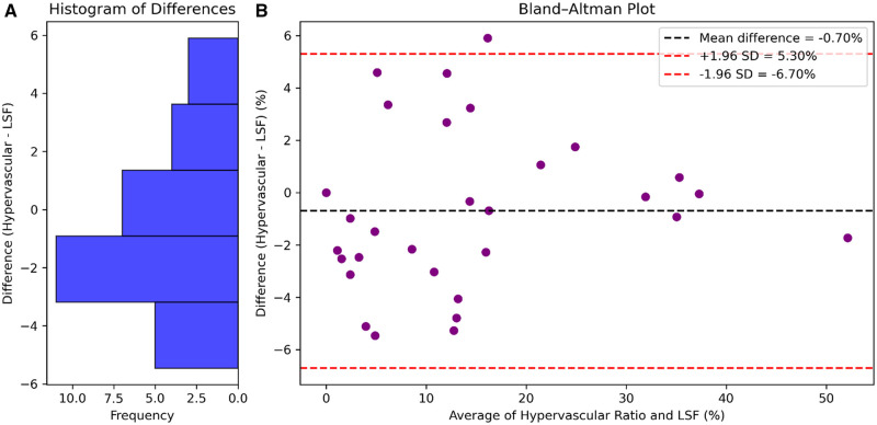
Results: The cohort consisted of 23 male and 7 female patients with a median age of 66 years (interquartile range: 58-71), diagnosed with hepatocellular carcinoma (n = 24), intrahepatic cholangiocarcinoma (n = 2), pancreatic neuroendocrine tumors (n = 2), metastatic colorectal cancer (n = 1), and lymphocyte carcinoma (n = 1). Regression of the hypervascular-tumor-to-perfused volume ratio on CECT against LSF from 99mTc-MAA imaging showed R2 = 0.95 (P < .001). In contrast, the correlation between tumor volume and LSF was R2 = 0.38 (P = .001). The root mean square error between the LSF estimated from CECT and that measured using 99mTc-MAA planar imaging was 3%.
Conclusion: Hypervascular-tumor-to-perfused volume ratio computed from CECT may offer a suitable alternative to 99mTc-MAA nuclear imaging for LSF estimation in patients undergoing transarterial radioembolization.

 求助内容:
求助内容: 应助结果提醒方式:


