Shawn K Lyo, Suyash Mohan, Michael J Hoch, Vivek P Patel, Robert M Kurtz, Alvand Hassankhani
{"title":"深度学习MRI使扫描时间减半,并在常规神经放射学检查中保持图像质量。","authors":"Shawn K Lyo, Suyash Mohan, Michael J Hoch, Vivek P Patel, Robert M Kurtz, Alvand Hassankhani","doi":"10.1093/radadv/umaf029","DOIUrl":null,"url":null,"abstract":"<p><strong>Background: </strong>Magnetic resonance imaging (MRI) is a cornerstone of neuroimaging but is limited by lengthy acquisition times, which can lead to motion artifacts, patient discomfort, and delayed care. Deep learning reconstruction is an emerging technology that can offer image acquisition acceleration while maintaining image quality.</p><p><strong>Purpose: </strong>To compare image quality and acquisition efficiency between deep learning-accelerated vs conventional MRI (C-MRI) across a spectrum of routine neuroradiologic examinations.</p><p><strong>Materials and methods: </strong>In this single-center retrospective study, 26 patients underwent imaging with a commercially available, FDA-cleared deep learning-accelerated MRI reconstruction algorithm (Deep Resolve, Siemens Healthineers), and C-MRI on a Siemens 3 T MAGNETOM Vida scanner between October 24 and November 14, 2023. A total of 113 sequence pairs were acquired across multiple body parts (brain [<i>n</i> = 28], cervical spine [<i>n</i> = 24], thoracic spine [<i>n </i>= 16], lumbar spine [<i>n</i> = 14], internal auditory canals [<i>n</i> = 5], sella [<i>n</i> = 5], neck [<i>n</i> = 5], temporomandibular joints [<i>n</i> = 6], brachial plexus [<i>n</i> = 4], and orbits [<i>n</i> = 6]) and sequences (T2 [<i>n</i> = 38], T1 [<i>n</i> = 30], short tau inversion recovery [<i>n</i> = 21], T1 post-contrast [<i>n</i> = 17], T2 fluid attenuated inversion recovery [<i>n</i> = 5], and proton density [<i>n </i>= 2]) and evaluated by 4 neuroradiologists blinded to the acquisition method for image quality using a 5-point Likert scale. Acquisition parameters were extracted from Digital Imaging and Communications in Medicine (DICOM) metadata and statistically compared. Rater preferences and interrater reliability were assessed using nonparametric tests and intraclass correlation coefficients.</p><p><strong>Results: </strong>Deep learning reduced mean scan time by 51.6% (95% CI: 45.7%-57.7%; from 110.8 seconds to 53.7 seconds; <i>P</i> < .001). Image quality assessments using a Likert scale showed scores slightly above neutral for signal-to-noise ratio (mean 3.51; 95% CI: 3.44-3.58), structural delineation (mean 3.51, 95% CI: 3.44-3.56), and overall image quality (mean 3.56, 95% CI: 3.49-3.63). However, poor interrater reliability (intraclass correlation [ICC] range: 0.06-0.33) showed that the observed differences were not consistent, indicating functional equivalence between conventional and deep learning images.</p><p><strong>Conclusion: </strong>Deep learning MRI enabled substantial scan time reductions while maintaining image quality.</p>","PeriodicalId":519940,"journal":{"name":"Radiology advances","volume":"2 5","pages":"umaf029"},"PeriodicalIF":0.0000,"publicationDate":"2025-08-23","publicationTypes":"Journal Article","fieldsOfStudy":null,"isOpenAccess":false,"openAccessPdf":"https://www.ncbi.nlm.nih.gov/pmc/articles/PMC12483150/pdf/","citationCount":"0","resultStr":"{\"title\":\"Deep learning MRI halves scan time and preserves image quality across routine neuroradiologic examinations.\",\"authors\":\"Shawn K Lyo, Suyash Mohan, Michael J Hoch, Vivek P Patel, Robert M Kurtz, Alvand Hassankhani\",\"doi\":\"10.1093/radadv/umaf029\",\"DOIUrl\":null,\"url\":null,\"abstract\":\"<p><strong>Background: </strong>Magnetic resonance imaging (MRI) is a cornerstone of neuroimaging but is limited by lengthy acquisition times, which can lead to motion artifacts, patient discomfort, and delayed care. Deep learning reconstruction is an emerging technology that can offer image acquisition acceleration while maintaining image quality.</p><p><strong>Purpose: </strong>To compare image quality and acquisition efficiency between deep learning-accelerated vs conventional MRI (C-MRI) across a spectrum of routine neuroradiologic examinations.</p><p><strong>Materials and methods: </strong>In this single-center retrospective study, 26 patients underwent imaging with a commercially available, FDA-cleared deep learning-accelerated MRI reconstruction algorithm (Deep Resolve, Siemens Healthineers), and C-MRI on a Siemens 3 T MAGNETOM Vida scanner between October 24 and November 14, 2023. A total of 113 sequence pairs were acquired across multiple body parts (brain [<i>n</i> = 28], cervical spine [<i>n</i> = 24], thoracic spine [<i>n </i>= 16], lumbar spine [<i>n</i> = 14], internal auditory canals [<i>n</i> = 5], sella [<i>n</i> = 5], neck [<i>n</i> = 5], temporomandibular joints [<i>n</i> = 6], brachial plexus [<i>n</i> = 4], and orbits [<i>n</i> = 6]) and sequences (T2 [<i>n</i> = 38], T1 [<i>n</i> = 30], short tau inversion recovery [<i>n</i> = 21], T1 post-contrast [<i>n</i> = 17], T2 fluid attenuated inversion recovery [<i>n</i> = 5], and proton density [<i>n </i>= 2]) and evaluated by 4 neuroradiologists blinded to the acquisition method for image quality using a 5-point Likert scale. Acquisition parameters were extracted from Digital Imaging and Communications in Medicine (DICOM) metadata and statistically compared. Rater preferences and interrater reliability were assessed using nonparametric tests and intraclass correlation coefficients.</p><p><strong>Results: </strong>Deep learning reduced mean scan time by 51.6% (95% CI: 45.7%-57.7%; from 110.8 seconds to 53.7 seconds; <i>P</i> < .001). Image quality assessments using a Likert scale showed scores slightly above neutral for signal-to-noise ratio (mean 3.51; 95% CI: 3.44-3.58), structural delineation (mean 3.51, 95% CI: 3.44-3.56), and overall image quality (mean 3.56, 95% CI: 3.49-3.63). However, poor interrater reliability (intraclass correlation [ICC] range: 0.06-0.33) showed that the observed differences were not consistent, indicating functional equivalence between conventional and deep learning images.</p><p><strong>Conclusion: </strong>Deep learning MRI enabled substantial scan time reductions while maintaining image quality.</p>\",\"PeriodicalId\":519940,\"journal\":{\"name\":\"Radiology advances\",\"volume\":\"2 5\",\"pages\":\"umaf029\"},\"PeriodicalIF\":0.0000,\"publicationDate\":\"2025-08-23\",\"publicationTypes\":\"Journal Article\",\"fieldsOfStudy\":null,\"isOpenAccess\":false,\"openAccessPdf\":\"https://www.ncbi.nlm.nih.gov/pmc/articles/PMC12483150/pdf/\",\"citationCount\":\"0\",\"resultStr\":null,\"platform\":\"Semanticscholar\",\"paperid\":null,\"PeriodicalName\":\"Radiology advances\",\"FirstCategoryId\":\"1085\",\"ListUrlMain\":\"https://doi.org/10.1093/radadv/umaf029\",\"RegionNum\":0,\"RegionCategory\":null,\"ArticlePicture\":[],\"TitleCN\":null,\"AbstractTextCN\":null,\"PMCID\":null,\"EPubDate\":\"2025/9/1 0:00:00\",\"PubModel\":\"eCollection\",\"JCR\":\"\",\"JCRName\":\"\",\"Score\":null,\"Total\":0}","platform":"Semanticscholar","paperid":null,"PeriodicalName":"Radiology advances","FirstCategoryId":"1085","ListUrlMain":"https://doi.org/10.1093/radadv/umaf029","RegionNum":0,"RegionCategory":null,"ArticlePicture":[],"TitleCN":null,"AbstractTextCN":null,"PMCID":null,"EPubDate":"2025/9/1 0:00:00","PubModel":"eCollection","JCR":"","JCRName":"","Score":null,"Total":0}
Deep learning MRI halves scan time and preserves image quality across routine neuroradiologic examinations.
Background: Magnetic resonance imaging (MRI) is a cornerstone of neuroimaging but is limited by lengthy acquisition times, which can lead to motion artifacts, patient discomfort, and delayed care. Deep learning reconstruction is an emerging technology that can offer image acquisition acceleration while maintaining image quality.
Purpose: To compare image quality and acquisition efficiency between deep learning-accelerated vs conventional MRI (C-MRI) across a spectrum of routine neuroradiologic examinations.
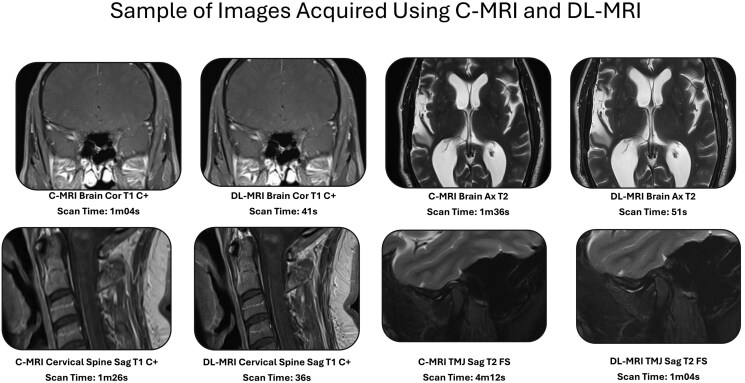
Materials and methods: In this single-center retrospective study, 26 patients underwent imaging with a commercially available, FDA-cleared deep learning-accelerated MRI reconstruction algorithm (Deep Resolve, Siemens Healthineers), and C-MRI on a Siemens 3 T MAGNETOM Vida scanner between October 24 and November 14, 2023. A total of 113 sequence pairs were acquired across multiple body parts (brain [n = 28], cervical spine [n = 24], thoracic spine [n = 16], lumbar spine [n = 14], internal auditory canals [n = 5], sella [n = 5], neck [n = 5], temporomandibular joints [n = 6], brachial plexus [n = 4], and orbits [n = 6]) and sequences (T2 [n = 38], T1 [n = 30], short tau inversion recovery [n = 21], T1 post-contrast [n = 17], T2 fluid attenuated inversion recovery [n = 5], and proton density [n = 2]) and evaluated by 4 neuroradiologists blinded to the acquisition method for image quality using a 5-point Likert scale. Acquisition parameters were extracted from Digital Imaging and Communications in Medicine (DICOM) metadata and statistically compared. Rater preferences and interrater reliability were assessed using nonparametric tests and intraclass correlation coefficients.
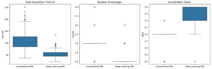
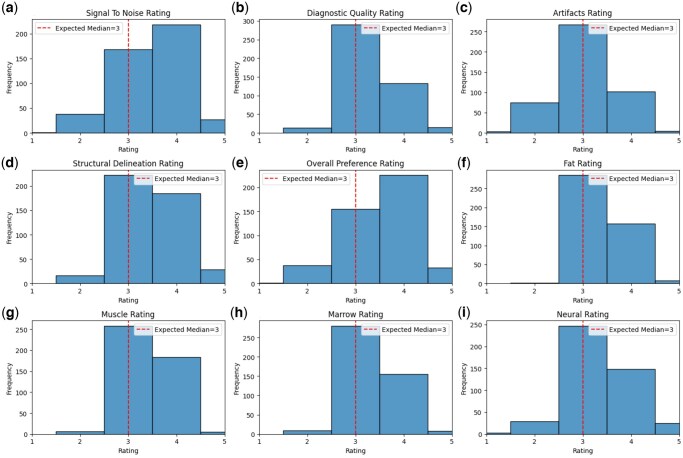
Results: Deep learning reduced mean scan time by 51.6% (95% CI: 45.7%-57.7%; from 110.8 seconds to 53.7 seconds; P < .001). Image quality assessments using a Likert scale showed scores slightly above neutral for signal-to-noise ratio (mean 3.51; 95% CI: 3.44-3.58), structural delineation (mean 3.51, 95% CI: 3.44-3.56), and overall image quality (mean 3.56, 95% CI: 3.49-3.63). However, poor interrater reliability (intraclass correlation [ICC] range: 0.06-0.33) showed that the observed differences were not consistent, indicating functional equivalence between conventional and deep learning images.
Conclusion: Deep learning MRI enabled substantial scan time reductions while maintaining image quality.

 求助内容:
求助内容: 应助结果提醒方式:


