Mina Chookhachizadeh Moghadam, Mohit Aspal, Xinzi He, Dominick J Romano, Arman Sharbatdaran, Zhongxiu Hu, Kurt Teichman, Hui Yi Ng He, Usama Sattar, Chenglin Zhu, Hreedi Dev, Daniil Shimonov, James M Chevalier, Akshay Goel, George Shih, Jon D Blumenfeld, Mert R Sabuncu, Martin R Prince
{"title":"基于深度学习的常染色体显性多囊肾病MRI肝囊肿分割。","authors":"Mina Chookhachizadeh Moghadam, Mohit Aspal, Xinzi He, Dominick J Romano, Arman Sharbatdaran, Zhongxiu Hu, Kurt Teichman, Hui Yi Ng He, Usama Sattar, Chenglin Zhu, Hreedi Dev, Daniil Shimonov, James M Chevalier, Akshay Goel, George Shih, Jon D Blumenfeld, Mert R Sabuncu, Martin R Prince","doi":"10.1093/radadv/umae014","DOIUrl":null,"url":null,"abstract":"<p><strong>Background: </strong>Autosomal dominant polycystic kidney disease (ADPKD) can lead to polycystic liver disease (PLD), characterized by liver cysts. Although majority of the patients are asymptomatic, massively enlarged liver secondary to PLD can cause discomfort, and compression on adjacent structures requiring cyst aspiration/fenestration, partial liver resection, or liver transplantation. Monitoring PLD by measuring liver volume fails to track the early stages when liver cyst volume is too small to affect liver volume.</p><p><strong>Purpose: </strong>To improve PLD assessment in the early stages by automating detection and segmentation of liver cysts using deep learning (DL) models.</p><p><strong>Materials and methods: </strong>A self-configured UNet-based platform (nnU-Net) was trained with 40 ADPKD subjects with liver cysts annotated by a radiologist. Internal (n = 7), External (n = 10), and test-retest reproducibility (n = 17) validations included macro- and micro-level performance metrics: patient-level Dice scores (PDice), along with voxel-level true positive rates (VTPR), as well as analysis of time saved in a model-assisted scenario. Additionally, we assessed human-level reliability in liver cyst segmentation and evaluated the model's test-retest reproducibility. We further compared liver volume vs cyst volume for tracking disease in a subject with 16+ years follow-up.</p><p><strong>Results: </strong>The model achieved an 82% ± 11% PDice and a 75% ± 15% VTPR on the internal test sets (n = 7 patients), and 80% ± 12% Dice score and a 91% ± 7% VTPR on the external test sets (n = 10 patients). It excelled particularly in detecting small liver cysts, a challenging task for manual annotation. This efficiency translated to a median of 91% (IQR: 14%) reduction in annotation time compared to manual labeling. Test-retest assessment demonstrated excellent reproducibility, with coefficients of variation of 94% for liver cyst fraction and 92% for cyst count.</p><p><strong>Conclusion: </strong>DL automation of liver cyst segmentations demonstrates potential to improve tracking of liver cyst volume in polycystic liver disease.</p>","PeriodicalId":519940,"journal":{"name":"Radiology advances","volume":"1 2","pages":"umae014"},"PeriodicalIF":0.0000,"publicationDate":"2024-05-23","publicationTypes":"Journal Article","fieldsOfStudy":null,"isOpenAccess":false,"openAccessPdf":"https://www.ncbi.nlm.nih.gov/pmc/articles/PMC12429238/pdf/","citationCount":"0","resultStr":"{\"title\":\"Deep learning-based liver cyst segmentation in MRI for autosomal dominant polycystic kidney disease.\",\"authors\":\"Mina Chookhachizadeh Moghadam, Mohit Aspal, Xinzi He, Dominick J Romano, Arman Sharbatdaran, Zhongxiu Hu, Kurt Teichman, Hui Yi Ng He, Usama Sattar, Chenglin Zhu, Hreedi Dev, Daniil Shimonov, James M Chevalier, Akshay Goel, George Shih, Jon D Blumenfeld, Mert R Sabuncu, Martin R Prince\",\"doi\":\"10.1093/radadv/umae014\",\"DOIUrl\":null,\"url\":null,\"abstract\":\"<p><strong>Background: </strong>Autosomal dominant polycystic kidney disease (ADPKD) can lead to polycystic liver disease (PLD), characterized by liver cysts. Although majority of the patients are asymptomatic, massively enlarged liver secondary to PLD can cause discomfort, and compression on adjacent structures requiring cyst aspiration/fenestration, partial liver resection, or liver transplantation. Monitoring PLD by measuring liver volume fails to track the early stages when liver cyst volume is too small to affect liver volume.</p><p><strong>Purpose: </strong>To improve PLD assessment in the early stages by automating detection and segmentation of liver cysts using deep learning (DL) models.</p><p><strong>Materials and methods: </strong>A self-configured UNet-based platform (nnU-Net) was trained with 40 ADPKD subjects with liver cysts annotated by a radiologist. Internal (n = 7), External (n = 10), and test-retest reproducibility (n = 17) validations included macro- and micro-level performance metrics: patient-level Dice scores (PDice), along with voxel-level true positive rates (VTPR), as well as analysis of time saved in a model-assisted scenario. Additionally, we assessed human-level reliability in liver cyst segmentation and evaluated the model's test-retest reproducibility. We further compared liver volume vs cyst volume for tracking disease in a subject with 16+ years follow-up.</p><p><strong>Results: </strong>The model achieved an 82% ± 11% PDice and a 75% ± 15% VTPR on the internal test sets (n = 7 patients), and 80% ± 12% Dice score and a 91% ± 7% VTPR on the external test sets (n = 10 patients). It excelled particularly in detecting small liver cysts, a challenging task for manual annotation. This efficiency translated to a median of 91% (IQR: 14%) reduction in annotation time compared to manual labeling. Test-retest assessment demonstrated excellent reproducibility, with coefficients of variation of 94% for liver cyst fraction and 92% for cyst count.</p><p><strong>Conclusion: </strong>DL automation of liver cyst segmentations demonstrates potential to improve tracking of liver cyst volume in polycystic liver disease.</p>\",\"PeriodicalId\":519940,\"journal\":{\"name\":\"Radiology advances\",\"volume\":\"1 2\",\"pages\":\"umae014\"},\"PeriodicalIF\":0.0000,\"publicationDate\":\"2024-05-23\",\"publicationTypes\":\"Journal Article\",\"fieldsOfStudy\":null,\"isOpenAccess\":false,\"openAccessPdf\":\"https://www.ncbi.nlm.nih.gov/pmc/articles/PMC12429238/pdf/\",\"citationCount\":\"0\",\"resultStr\":null,\"platform\":\"Semanticscholar\",\"paperid\":null,\"PeriodicalName\":\"Radiology advances\",\"FirstCategoryId\":\"1085\",\"ListUrlMain\":\"https://doi.org/10.1093/radadv/umae014\",\"RegionNum\":0,\"RegionCategory\":null,\"ArticlePicture\":[],\"TitleCN\":null,\"AbstractTextCN\":null,\"PMCID\":null,\"EPubDate\":\"2024/7/1 0:00:00\",\"PubModel\":\"eCollection\",\"JCR\":\"\",\"JCRName\":\"\",\"Score\":null,\"Total\":0}","platform":"Semanticscholar","paperid":null,"PeriodicalName":"Radiology advances","FirstCategoryId":"1085","ListUrlMain":"https://doi.org/10.1093/radadv/umae014","RegionNum":0,"RegionCategory":null,"ArticlePicture":[],"TitleCN":null,"AbstractTextCN":null,"PMCID":null,"EPubDate":"2024/7/1 0:00:00","PubModel":"eCollection","JCR":"","JCRName":"","Score":null,"Total":0}
Deep learning-based liver cyst segmentation in MRI for autosomal dominant polycystic kidney disease.
Background: Autosomal dominant polycystic kidney disease (ADPKD) can lead to polycystic liver disease (PLD), characterized by liver cysts. Although majority of the patients are asymptomatic, massively enlarged liver secondary to PLD can cause discomfort, and compression on adjacent structures requiring cyst aspiration/fenestration, partial liver resection, or liver transplantation. Monitoring PLD by measuring liver volume fails to track the early stages when liver cyst volume is too small to affect liver volume.
Purpose: To improve PLD assessment in the early stages by automating detection and segmentation of liver cysts using deep learning (DL) models.
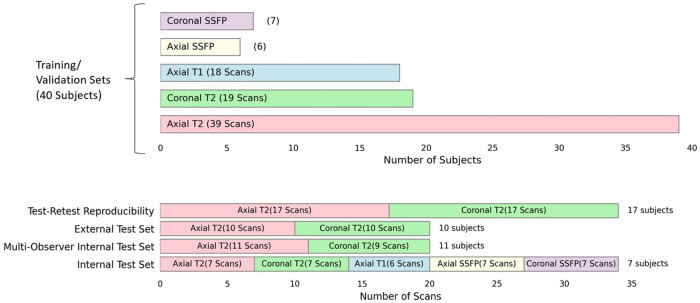
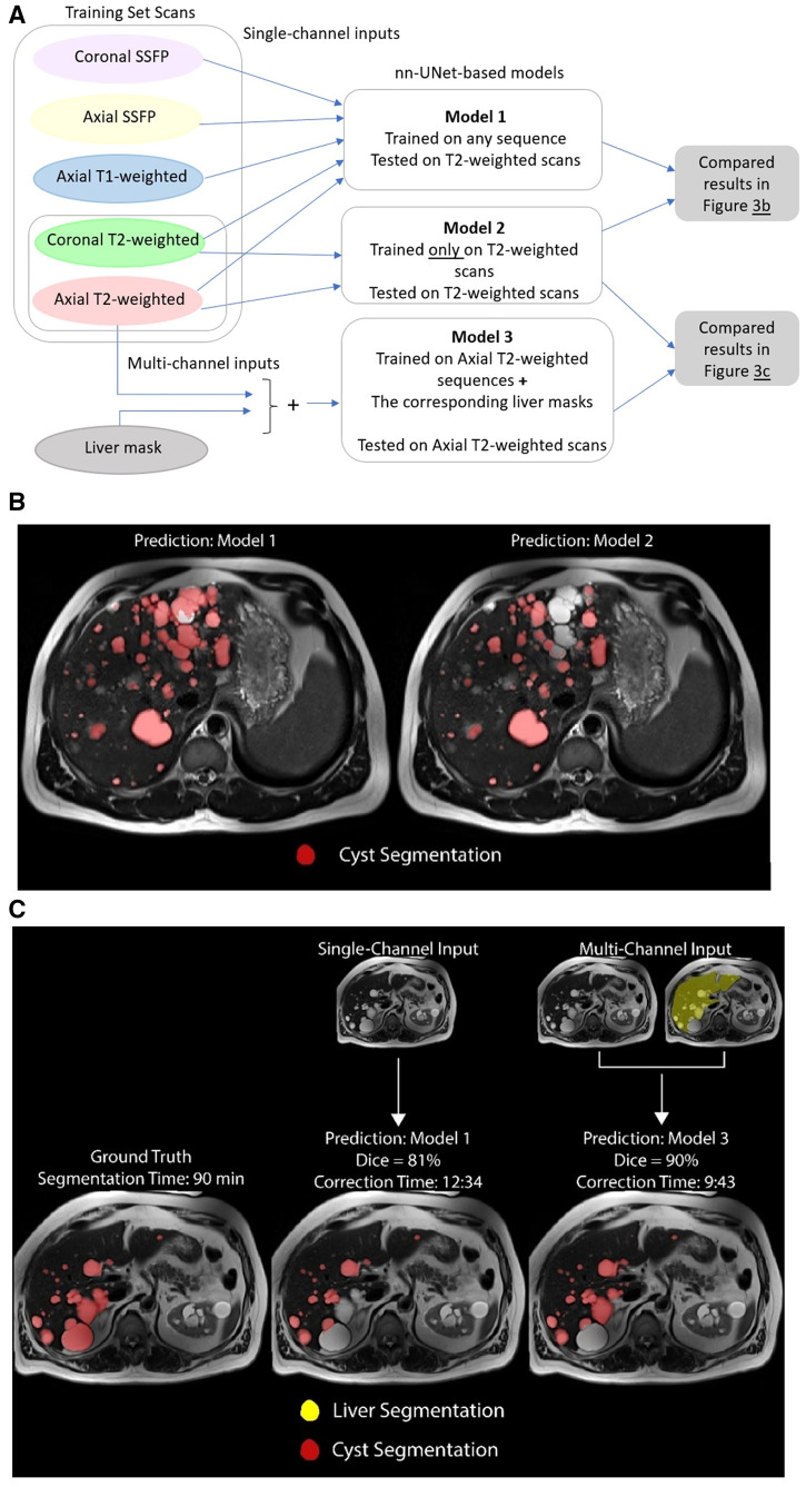
Materials and methods: A self-configured UNet-based platform (nnU-Net) was trained with 40 ADPKD subjects with liver cysts annotated by a radiologist. Internal (n = 7), External (n = 10), and test-retest reproducibility (n = 17) validations included macro- and micro-level performance metrics: patient-level Dice scores (PDice), along with voxel-level true positive rates (VTPR), as well as analysis of time saved in a model-assisted scenario. Additionally, we assessed human-level reliability in liver cyst segmentation and evaluated the model's test-retest reproducibility. We further compared liver volume vs cyst volume for tracking disease in a subject with 16+ years follow-up.
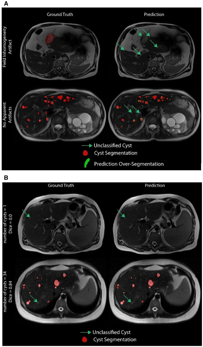
Results: The model achieved an 82% ± 11% PDice and a 75% ± 15% VTPR on the internal test sets (n = 7 patients), and 80% ± 12% Dice score and a 91% ± 7% VTPR on the external test sets (n = 10 patients). It excelled particularly in detecting small liver cysts, a challenging task for manual annotation. This efficiency translated to a median of 91% (IQR: 14%) reduction in annotation time compared to manual labeling. Test-retest assessment demonstrated excellent reproducibility, with coefficients of variation of 94% for liver cyst fraction and 92% for cyst count.
Conclusion: DL automation of liver cyst segmentations demonstrates potential to improve tracking of liver cyst volume in polycystic liver disease.

 求助内容:
求助内容: 应助结果提醒方式:


