Matthias Schwab, Mathias Pamminger, Christian Kremser, Markus Haltmeier, Agnes Mayr
{"title":"基于临床心脏MR扫描的全自动心肌梗死分割的深度学习管道。","authors":"Matthias Schwab, Mathias Pamminger, Christian Kremser, Markus Haltmeier, Agnes Mayr","doi":"10.1093/radadv/umaf023","DOIUrl":null,"url":null,"abstract":"<p><strong>Background: </strong>Artificial intelligence (AI) has demonstrated promise in cardiovascular magnetic resonance (CMR) imaging, particularly in myocardial infarct segmentation, where it may help reduce variability and workload in clinical practice.</p><p><strong>Purpose: </strong>To develop and evaluate a deep learning-based model that performs myocardial infarct segmentation in a fully automated way.</p><p><strong>Materials and methods: </strong>For this retrospective study, a cascaded framework of 2- and 3-dimensional convolutional neural networks (CNNs), specialized in identifying ischemic myocardial scars on late gadolinium enhancement (LGE) CMR images, was trained on an in-house training dataset of 144 examinations acquired using a 1.5 Tesla Siemens scanner collected between 2006 and 2022. On a separate test dataset from the same institution, comprising images from 152 examinations, a quantitative comparison was conducted between AI-based segmentations and manual segmentations. Further, segmentation accuracy was assessed qualitatively for both human and AI-generated contours by 2 CMR experts in a blinded experiment. Most cases underwent single human assessment, with double reading conducted only on a subset of 20 cases.</p><p><strong>Results: </strong>Excellent agreement was found between manually and automatically calculated infarct volumes (ρ<sub>c</sub> = 0.9). The qualitative evaluation showed that compared to human-based measurements, the experts rated the AI-based segmentations as better representing the actual extent of infarction (<i>P</i> < 0.001) and preferred them more often (33.4% AI, 25.1% human, 41.5% equal). On the contrary, for segmentation of microvascular obstruction (MVO), manual measurements were still preferred (<i>P</i> < 0.001; 11.3% AI, 55.6% human, 33.1% equal).</p><p><strong>Conclusion: </strong>This fully automated segmentation pipeline enables the calculation of CMR infarct size without requiring any pre-processing of the input images while matching the segmentation quality of trained human observers. As automated infarct segmentation is preferred over manual segmentation, further development of this workflow toward clinical application is warranted to improve efficiencies.</p><p><strong>Summary: </strong>We developed and evaluated an algorithm that performs myocardial infarct segmentation from cardiac MR images without requiring pre-processing and that outperforms trained human observers on qualitative expert judgment.</p>","PeriodicalId":519940,"journal":{"name":"Radiology advances","volume":"2 4","pages":"umaf023"},"PeriodicalIF":0.0000,"publicationDate":"2025-07-18","publicationTypes":"Journal Article","fieldsOfStudy":null,"isOpenAccess":false,"openAccessPdf":"https://www.ncbi.nlm.nih.gov/pmc/articles/PMC12429236/pdf/","citationCount":"0","resultStr":"{\"title\":\"Deep learning pipeline for fully automated myocardial infarct segmentation from clinical cardiac MR scans.\",\"authors\":\"Matthias Schwab, Mathias Pamminger, Christian Kremser, Markus Haltmeier, Agnes Mayr\",\"doi\":\"10.1093/radadv/umaf023\",\"DOIUrl\":null,\"url\":null,\"abstract\":\"<p><strong>Background: </strong>Artificial intelligence (AI) has demonstrated promise in cardiovascular magnetic resonance (CMR) imaging, particularly in myocardial infarct segmentation, where it may help reduce variability and workload in clinical practice.</p><p><strong>Purpose: </strong>To develop and evaluate a deep learning-based model that performs myocardial infarct segmentation in a fully automated way.</p><p><strong>Materials and methods: </strong>For this retrospective study, a cascaded framework of 2- and 3-dimensional convolutional neural networks (CNNs), specialized in identifying ischemic myocardial scars on late gadolinium enhancement (LGE) CMR images, was trained on an in-house training dataset of 144 examinations acquired using a 1.5 Tesla Siemens scanner collected between 2006 and 2022. On a separate test dataset from the same institution, comprising images from 152 examinations, a quantitative comparison was conducted between AI-based segmentations and manual segmentations. Further, segmentation accuracy was assessed qualitatively for both human and AI-generated contours by 2 CMR experts in a blinded experiment. Most cases underwent single human assessment, with double reading conducted only on a subset of 20 cases.</p><p><strong>Results: </strong>Excellent agreement was found between manually and automatically calculated infarct volumes (ρ<sub>c</sub> = 0.9). The qualitative evaluation showed that compared to human-based measurements, the experts rated the AI-based segmentations as better representing the actual extent of infarction (<i>P</i> < 0.001) and preferred them more often (33.4% AI, 25.1% human, 41.5% equal). On the contrary, for segmentation of microvascular obstruction (MVO), manual measurements were still preferred (<i>P</i> < 0.001; 11.3% AI, 55.6% human, 33.1% equal).</p><p><strong>Conclusion: </strong>This fully automated segmentation pipeline enables the calculation of CMR infarct size without requiring any pre-processing of the input images while matching the segmentation quality of trained human observers. As automated infarct segmentation is preferred over manual segmentation, further development of this workflow toward clinical application is warranted to improve efficiencies.</p><p><strong>Summary: </strong>We developed and evaluated an algorithm that performs myocardial infarct segmentation from cardiac MR images without requiring pre-processing and that outperforms trained human observers on qualitative expert judgment.</p>\",\"PeriodicalId\":519940,\"journal\":{\"name\":\"Radiology advances\",\"volume\":\"2 4\",\"pages\":\"umaf023\"},\"PeriodicalIF\":0.0000,\"publicationDate\":\"2025-07-18\",\"publicationTypes\":\"Journal Article\",\"fieldsOfStudy\":null,\"isOpenAccess\":false,\"openAccessPdf\":\"https://www.ncbi.nlm.nih.gov/pmc/articles/PMC12429236/pdf/\",\"citationCount\":\"0\",\"resultStr\":null,\"platform\":\"Semanticscholar\",\"paperid\":null,\"PeriodicalName\":\"Radiology advances\",\"FirstCategoryId\":\"1085\",\"ListUrlMain\":\"https://doi.org/10.1093/radadv/umaf023\",\"RegionNum\":0,\"RegionCategory\":null,\"ArticlePicture\":[],\"TitleCN\":null,\"AbstractTextCN\":null,\"PMCID\":null,\"EPubDate\":\"2025/7/1 0:00:00\",\"PubModel\":\"eCollection\",\"JCR\":\"\",\"JCRName\":\"\",\"Score\":null,\"Total\":0}","platform":"Semanticscholar","paperid":null,"PeriodicalName":"Radiology advances","FirstCategoryId":"1085","ListUrlMain":"https://doi.org/10.1093/radadv/umaf023","RegionNum":0,"RegionCategory":null,"ArticlePicture":[],"TitleCN":null,"AbstractTextCN":null,"PMCID":null,"EPubDate":"2025/7/1 0:00:00","PubModel":"eCollection","JCR":"","JCRName":"","Score":null,"Total":0}
Deep learning pipeline for fully automated myocardial infarct segmentation from clinical cardiac MR scans.
Background: Artificial intelligence (AI) has demonstrated promise in cardiovascular magnetic resonance (CMR) imaging, particularly in myocardial infarct segmentation, where it may help reduce variability and workload in clinical practice.
Purpose: To develop and evaluate a deep learning-based model that performs myocardial infarct segmentation in a fully automated way.
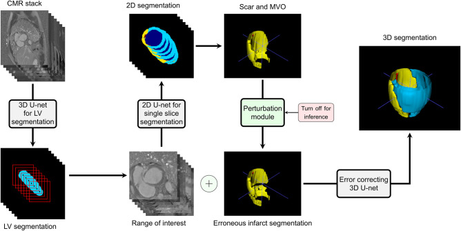
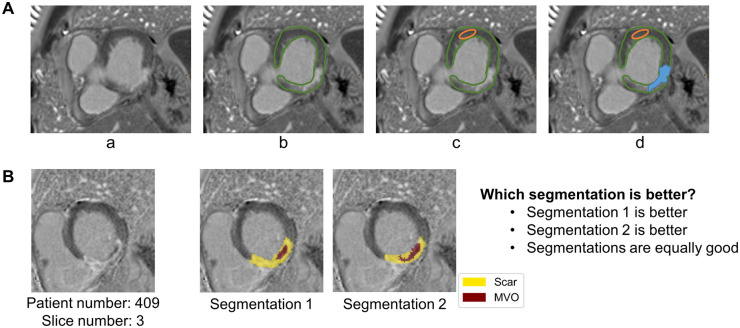
Materials and methods: For this retrospective study, a cascaded framework of 2- and 3-dimensional convolutional neural networks (CNNs), specialized in identifying ischemic myocardial scars on late gadolinium enhancement (LGE) CMR images, was trained on an in-house training dataset of 144 examinations acquired using a 1.5 Tesla Siemens scanner collected between 2006 and 2022. On a separate test dataset from the same institution, comprising images from 152 examinations, a quantitative comparison was conducted between AI-based segmentations and manual segmentations. Further, segmentation accuracy was assessed qualitatively for both human and AI-generated contours by 2 CMR experts in a blinded experiment. Most cases underwent single human assessment, with double reading conducted only on a subset of 20 cases.
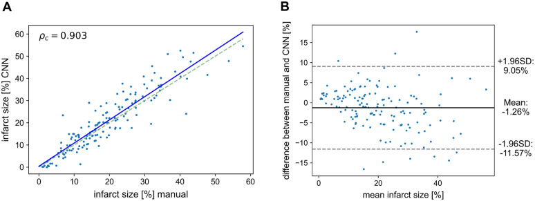
Results: Excellent agreement was found between manually and automatically calculated infarct volumes (ρc = 0.9). The qualitative evaluation showed that compared to human-based measurements, the experts rated the AI-based segmentations as better representing the actual extent of infarction (P < 0.001) and preferred them more often (33.4% AI, 25.1% human, 41.5% equal). On the contrary, for segmentation of microvascular obstruction (MVO), manual measurements were still preferred (P < 0.001; 11.3% AI, 55.6% human, 33.1% equal).
Conclusion: This fully automated segmentation pipeline enables the calculation of CMR infarct size without requiring any pre-processing of the input images while matching the segmentation quality of trained human observers. As automated infarct segmentation is preferred over manual segmentation, further development of this workflow toward clinical application is warranted to improve efficiencies.
Summary: We developed and evaluated an algorithm that performs myocardial infarct segmentation from cardiac MR images without requiring pre-processing and that outperforms trained human observers on qualitative expert judgment.

 求助内容:
求助内容: 应助结果提醒方式:


