Colin C. Gauvin, , , Monika Tokmina-Lukaszewska, , , Hitesh Kumar Waghwani, , , Sterling C. McBee, , , Trevor Douglas, , , Brian Bothner, , and , C. Martin Lawrence*,
{"title":"微型铁蛋白中矿物成核和生长的机制","authors":"Colin C. Gauvin, , , Monika Tokmina-Lukaszewska, , , Hitesh Kumar Waghwani, , , Sterling C. McBee, , , Trevor Douglas, , , Brian Bothner, , and , C. Martin Lawrence*, ","doi":"10.1021/jacs.5c05464","DOIUrl":null,"url":null,"abstract":"<p >Iron is an enigmatic element. While necessary for life, as Fe(II) it also catalyzes formation of reactive oxygen species. To mitigate this, cellular life has evolved the ferritin protein superfamily, which includes the 24 subunit ferritins and bacterioferritins, and 12 subunit mini-ferritins (DPS). Each catalyze the oxidation of Fe(II) to ferric oxyhydroxide, which is then sequestered within the hollow protein shell. While there is a wealth of structural information on unmineralized ferritins, high resolution information on iron loaded ferritins is lacking, and the mechanism of iron mineralization is poorly understood. To address this, we followed iron loading in a mini-ferritin by cryo-EM. We determined a 1.86 Å structure in the unmineralized state, as well as a 1.91 Å structure of an early, iron loading state in which the mini-ferritin catalyzes nucleation of ferric oxyhydroxide at the acidic 3-fold pores. Mechanistically, a conserved crucible of precisely positioned glutamates and unsaturated main chain carbonyls are employed as a template to catalyze nucleation. A 2.4 Å structure at a later time point was also determined, revealing the role of a second constellation of main-chain carbonyls on the interior surface that subsequently supports crystalline mineral growth, that then proceeds into the center of the particle. Notably, the visualized mineral is consistent with one of two competing structural descriptions for ferrihydrite. This study provides the first pseudoatomic level observation of controlled mineral nucleation and growth in any member of the ferritin superfamily, and informs general mechanisms of nucleation and biomineralization.</p>","PeriodicalId":49,"journal":{"name":"Journal of the American Chemical Society","volume":"147 41","pages":"37030–37044"},"PeriodicalIF":15.6000,"publicationDate":"2025-10-06","publicationTypes":"Journal Article","fieldsOfStudy":null,"isOpenAccess":false,"openAccessPdf":"https://pubs.acs.org/doi/pdf/10.1021/jacs.5c05464","citationCount":"0","resultStr":"{\"title\":\"The Mechanism of Mineral Nucleation and Growth in a Mini-Ferritin\",\"authors\":\"Colin C. Gauvin, , , Monika Tokmina-Lukaszewska, , , Hitesh Kumar Waghwani, , , Sterling C. McBee, , , Trevor Douglas, , , Brian Bothner, , and , C. Martin Lawrence*, \",\"doi\":\"10.1021/jacs.5c05464\",\"DOIUrl\":null,\"url\":null,\"abstract\":\"<p >Iron is an enigmatic element. While necessary for life, as Fe(II) it also catalyzes formation of reactive oxygen species. To mitigate this, cellular life has evolved the ferritin protein superfamily, which includes the 24 subunit ferritins and bacterioferritins, and 12 subunit mini-ferritins (DPS). Each catalyze the oxidation of Fe(II) to ferric oxyhydroxide, which is then sequestered within the hollow protein shell. While there is a wealth of structural information on unmineralized ferritins, high resolution information on iron loaded ferritins is lacking, and the mechanism of iron mineralization is poorly understood. To address this, we followed iron loading in a mini-ferritin by cryo-EM. We determined a 1.86 Å structure in the unmineralized state, as well as a 1.91 Å structure of an early, iron loading state in which the mini-ferritin catalyzes nucleation of ferric oxyhydroxide at the acidic 3-fold pores. Mechanistically, a conserved crucible of precisely positioned glutamates and unsaturated main chain carbonyls are employed as a template to catalyze nucleation. A 2.4 Å structure at a later time point was also determined, revealing the role of a second constellation of main-chain carbonyls on the interior surface that subsequently supports crystalline mineral growth, that then proceeds into the center of the particle. Notably, the visualized mineral is consistent with one of two competing structural descriptions for ferrihydrite. This study provides the first pseudoatomic level observation of controlled mineral nucleation and growth in any member of the ferritin superfamily, and informs general mechanisms of nucleation and biomineralization.</p>\",\"PeriodicalId\":49,\"journal\":{\"name\":\"Journal of the American Chemical Society\",\"volume\":\"147 41\",\"pages\":\"37030–37044\"},\"PeriodicalIF\":15.6000,\"publicationDate\":\"2025-10-06\",\"publicationTypes\":\"Journal Article\",\"fieldsOfStudy\":null,\"isOpenAccess\":false,\"openAccessPdf\":\"https://pubs.acs.org/doi/pdf/10.1021/jacs.5c05464\",\"citationCount\":\"0\",\"resultStr\":null,\"platform\":\"Semanticscholar\",\"paperid\":null,\"PeriodicalName\":\"Journal of the American Chemical Society\",\"FirstCategoryId\":\"92\",\"ListUrlMain\":\"https://pubs.acs.org/doi/10.1021/jacs.5c05464\",\"RegionNum\":1,\"RegionCategory\":\"化学\",\"ArticlePicture\":[],\"TitleCN\":null,\"AbstractTextCN\":null,\"PMCID\":null,\"EPubDate\":\"\",\"PubModel\":\"\",\"JCR\":\"Q1\",\"JCRName\":\"CHEMISTRY, MULTIDISCIPLINARY\",\"Score\":null,\"Total\":0}","platform":"Semanticscholar","paperid":null,"PeriodicalName":"Journal of the American Chemical Society","FirstCategoryId":"92","ListUrlMain":"https://pubs.acs.org/doi/10.1021/jacs.5c05464","RegionNum":1,"RegionCategory":"化学","ArticlePicture":[],"TitleCN":null,"AbstractTextCN":null,"PMCID":null,"EPubDate":"","PubModel":"","JCR":"Q1","JCRName":"CHEMISTRY, MULTIDISCIPLINARY","Score":null,"Total":0}
The Mechanism of Mineral Nucleation and Growth in a Mini-Ferritin
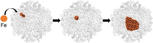
Iron is an enigmatic element. While necessary for life, as Fe(II) it also catalyzes formation of reactive oxygen species. To mitigate this, cellular life has evolved the ferritin protein superfamily, which includes the 24 subunit ferritins and bacterioferritins, and 12 subunit mini-ferritins (DPS). Each catalyze the oxidation of Fe(II) to ferric oxyhydroxide, which is then sequestered within the hollow protein shell. While there is a wealth of structural information on unmineralized ferritins, high resolution information on iron loaded ferritins is lacking, and the mechanism of iron mineralization is poorly understood. To address this, we followed iron loading in a mini-ferritin by cryo-EM. We determined a 1.86 Å structure in the unmineralized state, as well as a 1.91 Å structure of an early, iron loading state in which the mini-ferritin catalyzes nucleation of ferric oxyhydroxide at the acidic 3-fold pores. Mechanistically, a conserved crucible of precisely positioned glutamates and unsaturated main chain carbonyls are employed as a template to catalyze nucleation. A 2.4 Å structure at a later time point was also determined, revealing the role of a second constellation of main-chain carbonyls on the interior surface that subsequently supports crystalline mineral growth, that then proceeds into the center of the particle. Notably, the visualized mineral is consistent with one of two competing structural descriptions for ferrihydrite. This study provides the first pseudoatomic level observation of controlled mineral nucleation and growth in any member of the ferritin superfamily, and informs general mechanisms of nucleation and biomineralization.
期刊介绍:
The flagship journal of the American Chemical Society, known as the Journal of the American Chemical Society (JACS), has been a prestigious publication since its establishment in 1879. It holds a preeminent position in the field of chemistry and related interdisciplinary sciences. JACS is committed to disseminating cutting-edge research papers, covering a wide range of topics, and encompasses approximately 19,000 pages of Articles, Communications, and Perspectives annually. With a weekly publication frequency, JACS plays a vital role in advancing the field of chemistry by providing essential research.
 求助内容:
求助内容: 应助结果提醒方式:


