Zhen-Yu Li, Jing-Hua Yue, Wen-Jie Wu, Bo Liu, Jie Zhang
{"title":"基于深度学习的腮腺癌术后近距离放疗靶体积自动分割模型。","authors":"Zhen-Yu Li, Jing-Hua Yue, Wen-Jie Wu, Bo Liu, Jie Zhang","doi":"10.5114/jcb.2025.153913","DOIUrl":null,"url":null,"abstract":"<p><strong>Purpose: </strong>Timely and accurate delineation of the clinical target volume (CTV) in brachytherapy after parotid cancer surgery plays a crucial role in tailored delivery of radiation doses. This study aimed to develop and evaluate a deep learning-based model for auto-segmentation of the CTVs in postoperative adjuvant brachytherapy for patients with parotid gland cancer, addressing the challenge of achieving consistent, high-quality CTV delineations efficiently.</p><p><strong>Material and methods: </strong>Using clinical imaging data from 326 patients with parotid gland carcinoma treated at Peking University School and Hospital of Stomatology between 2017 and 2023, we established a training dataset of 213 cases, a validation set of 53 cases, and a test set of 60 cases. The CTVs on the images were segmented using 3D Res-UNet, a deep learning model, and compared against manual delineations performed by experienced radiation oncologists. The performance of 3D Res-UNet was optimized through a comprehensive preprocessing and training process tailored to the dataset's characteristics.</p><p><strong>Results: </strong>The deep learning model yielded a significant improvement in segmentation efficiency. The deep learning model generated initial CTV contours in 9.4 seconds of computational time. Subsequent expert review and minor adjustments required an average of 11.9 minutes, substantially shorter than the 46.7 minutes needed for fully manual delineation. Quantitative analysis showed that the Dice similarity coefficient (DSC) of automatic segmentation by 3D Res-UNet was 0.709, which improved to 0.924 after expert review. Qualitative evaluation by senior oncologists further affirmed the clinical acceptability of the automatically segmented CTVs.</p><p><strong>Conclusions: </strong>Automatic contouring with physician review enabled high-accuracy and rapid CTV generation, reducing the overall delineation workload by more than 30 minutes. Consequently, the proposed deep-learning model functions as a useful support tool that streamlines postoperative adjuvant brachytherapy planning for parotid gland cancer and lessens the burden on radiation oncologists, thereby contributing to improved patient care.</p>","PeriodicalId":51305,"journal":{"name":"Journal of Contemporary Brachytherapy","volume":"17 4","pages":"232-241"},"PeriodicalIF":1.1000,"publicationDate":"2025-08-01","publicationTypes":"Journal Article","fieldsOfStudy":null,"isOpenAccess":false,"openAccessPdf":"https://www.ncbi.nlm.nih.gov/pmc/articles/PMC12489543/pdf/","citationCount":"0","resultStr":"{\"title\":\"Deep learning-based auto-segmentation model for clinical target volume delineation in brachytherapy after parotid cancer surgery.\",\"authors\":\"Zhen-Yu Li, Jing-Hua Yue, Wen-Jie Wu, Bo Liu, Jie Zhang\",\"doi\":\"10.5114/jcb.2025.153913\",\"DOIUrl\":null,\"url\":null,\"abstract\":\"<p><strong>Purpose: </strong>Timely and accurate delineation of the clinical target volume (CTV) in brachytherapy after parotid cancer surgery plays a crucial role in tailored delivery of radiation doses. This study aimed to develop and evaluate a deep learning-based model for auto-segmentation of the CTVs in postoperative adjuvant brachytherapy for patients with parotid gland cancer, addressing the challenge of achieving consistent, high-quality CTV delineations efficiently.</p><p><strong>Material and methods: </strong>Using clinical imaging data from 326 patients with parotid gland carcinoma treated at Peking University School and Hospital of Stomatology between 2017 and 2023, we established a training dataset of 213 cases, a validation set of 53 cases, and a test set of 60 cases. The CTVs on the images were segmented using 3D Res-UNet, a deep learning model, and compared against manual delineations performed by experienced radiation oncologists. The performance of 3D Res-UNet was optimized through a comprehensive preprocessing and training process tailored to the dataset's characteristics.</p><p><strong>Results: </strong>The deep learning model yielded a significant improvement in segmentation efficiency. The deep learning model generated initial CTV contours in 9.4 seconds of computational time. Subsequent expert review and minor adjustments required an average of 11.9 minutes, substantially shorter than the 46.7 minutes needed for fully manual delineation. Quantitative analysis showed that the Dice similarity coefficient (DSC) of automatic segmentation by 3D Res-UNet was 0.709, which improved to 0.924 after expert review. Qualitative evaluation by senior oncologists further affirmed the clinical acceptability of the automatically segmented CTVs.</p><p><strong>Conclusions: </strong>Automatic contouring with physician review enabled high-accuracy and rapid CTV generation, reducing the overall delineation workload by more than 30 minutes. Consequently, the proposed deep-learning model functions as a useful support tool that streamlines postoperative adjuvant brachytherapy planning for parotid gland cancer and lessens the burden on radiation oncologists, thereby contributing to improved patient care.</p>\",\"PeriodicalId\":51305,\"journal\":{\"name\":\"Journal of Contemporary Brachytherapy\",\"volume\":\"17 4\",\"pages\":\"232-241\"},\"PeriodicalIF\":1.1000,\"publicationDate\":\"2025-08-01\",\"publicationTypes\":\"Journal Article\",\"fieldsOfStudy\":null,\"isOpenAccess\":false,\"openAccessPdf\":\"https://www.ncbi.nlm.nih.gov/pmc/articles/PMC12489543/pdf/\",\"citationCount\":\"0\",\"resultStr\":null,\"platform\":\"Semanticscholar\",\"paperid\":null,\"PeriodicalName\":\"Journal of Contemporary Brachytherapy\",\"FirstCategoryId\":\"3\",\"ListUrlMain\":\"https://doi.org/10.5114/jcb.2025.153913\",\"RegionNum\":4,\"RegionCategory\":\"医学\",\"ArticlePicture\":[],\"TitleCN\":null,\"AbstractTextCN\":null,\"PMCID\":null,\"EPubDate\":\"2025/8/28 0:00:00\",\"PubModel\":\"Epub\",\"JCR\":\"Q4\",\"JCRName\":\"ONCOLOGY\",\"Score\":null,\"Total\":0}","platform":"Semanticscholar","paperid":null,"PeriodicalName":"Journal of Contemporary Brachytherapy","FirstCategoryId":"3","ListUrlMain":"https://doi.org/10.5114/jcb.2025.153913","RegionNum":4,"RegionCategory":"医学","ArticlePicture":[],"TitleCN":null,"AbstractTextCN":null,"PMCID":null,"EPubDate":"2025/8/28 0:00:00","PubModel":"Epub","JCR":"Q4","JCRName":"ONCOLOGY","Score":null,"Total":0}
Deep learning-based auto-segmentation model for clinical target volume delineation in brachytherapy after parotid cancer surgery.
Purpose: Timely and accurate delineation of the clinical target volume (CTV) in brachytherapy after parotid cancer surgery plays a crucial role in tailored delivery of radiation doses. This study aimed to develop and evaluate a deep learning-based model for auto-segmentation of the CTVs in postoperative adjuvant brachytherapy for patients with parotid gland cancer, addressing the challenge of achieving consistent, high-quality CTV delineations efficiently.
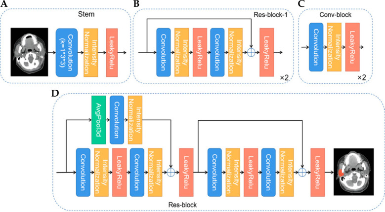
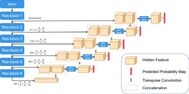
Material and methods: Using clinical imaging data from 326 patients with parotid gland carcinoma treated at Peking University School and Hospital of Stomatology between 2017 and 2023, we established a training dataset of 213 cases, a validation set of 53 cases, and a test set of 60 cases. The CTVs on the images were segmented using 3D Res-UNet, a deep learning model, and compared against manual delineations performed by experienced radiation oncologists. The performance of 3D Res-UNet was optimized through a comprehensive preprocessing and training process tailored to the dataset's characteristics.
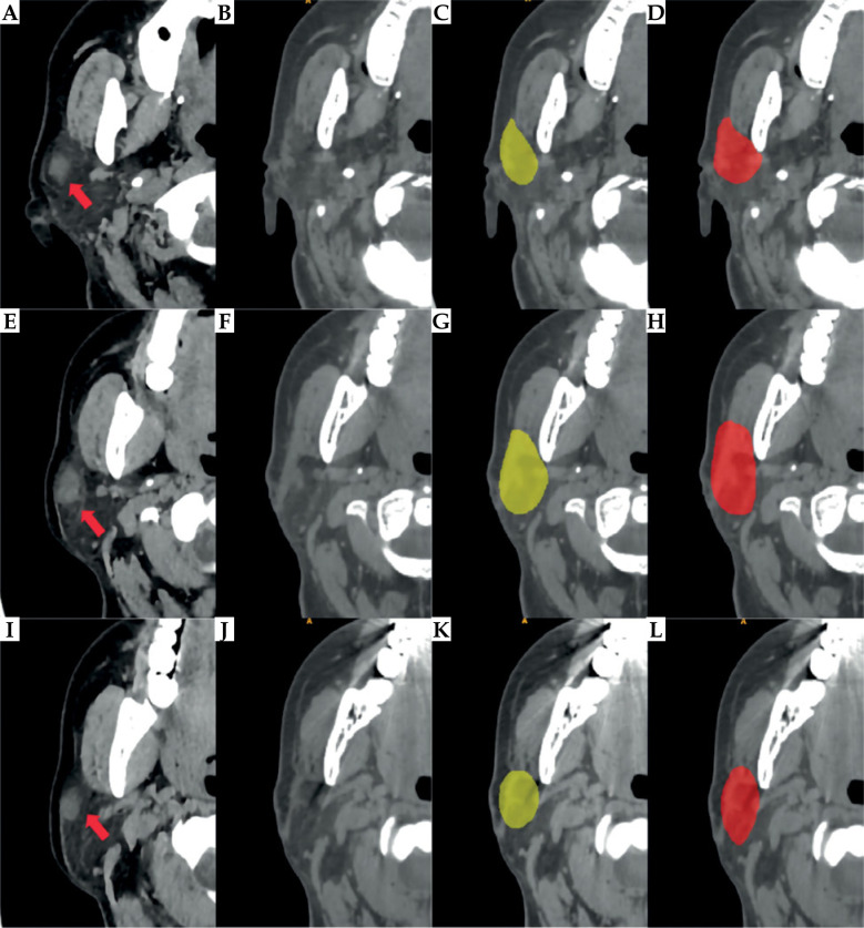
Results: The deep learning model yielded a significant improvement in segmentation efficiency. The deep learning model generated initial CTV contours in 9.4 seconds of computational time. Subsequent expert review and minor adjustments required an average of 11.9 minutes, substantially shorter than the 46.7 minutes needed for fully manual delineation. Quantitative analysis showed that the Dice similarity coefficient (DSC) of automatic segmentation by 3D Res-UNet was 0.709, which improved to 0.924 after expert review. Qualitative evaluation by senior oncologists further affirmed the clinical acceptability of the automatically segmented CTVs.
Conclusions: Automatic contouring with physician review enabled high-accuracy and rapid CTV generation, reducing the overall delineation workload by more than 30 minutes. Consequently, the proposed deep-learning model functions as a useful support tool that streamlines postoperative adjuvant brachytherapy planning for parotid gland cancer and lessens the burden on radiation oncologists, thereby contributing to improved patient care.
期刊介绍:
The “Journal of Contemporary Brachytherapy” is an international and multidisciplinary journal that will publish papers of original research as well as reviews of articles. Main subjects of the journal include: clinical brachytherapy, combined modality treatment, advances in radiobiology, hyperthermia and tumour biology, as well as physical aspects relevant to brachytherapy, particularly in the field of imaging, dosimetry and radiation therapy planning. Original contributions will include experimental studies of combined modality treatment, tumor sensitization and normal tissue protection, molecular radiation biology, and clinical investigations of cancer treatment in brachytherapy. Another field of interest will be the educational part of the journal.
 求助内容:
求助内容: 应助结果提醒方式:


