Lei Yan, Yang Liu, Yu-Yu Yan, Wei Li, Hong-Wei Li, Yun-Long Wang, Zhen-Fang Zhang, Xiao-Ling Li
{"title":"生物功能修复系统设计的下颌全口义齿模型修正区分析。","authors":"Lei Yan, Yang Liu, Yu-Yu Yan, Wei Li, Hong-Wei Li, Yun-Long Wang, Zhen-Fang Zhang, Xiao-Ling Li","doi":"10.2147/TCRM.S540674","DOIUrl":null,"url":null,"abstract":"<p><strong>Objective: </strong>This study aimed to analyze the regions and extent of modifications made to the final mandibular complete denture model designed using the bio-functional prosthetic system (BPS), compared to the initial model, utilizing digital scanning technology.</p><p><strong>Methods: </strong>Twenty individuals with edentulism requiring mandibular restoration using BPS were included in the study. Digital scans of the initial and final gypsum models of the edentulous mandible were obtained. The models were aligned, registered, and analyzed for modification zones using Geomagic Warp software.</p><p><strong>Results: </strong>Statistically significant differences were observed in three specific regions: the posterior fossa of the mylohyoid, the submandibular gland region, and the labial vestibule (<i>p</i> < 0.05). In contrast, differences in the posterior molar pad area were minimal (<i>p</i> > 0.05). Overall, the final model exhibited significant deviations from the initial model (<i>p</i> < 0.05), with the greatest variation observed in the posterior fossa of the mylohyoid and the least variation in the posterior molar pad area.</p><p><strong>Conclusion: </strong>The final mandibular complete denture model, constructed using BPS, demonstrates improved accuracy in representing mucosal movement, shape, and positioning under occlusal pressure compared to the initial model. The posterior fossa of the mylohyoid, submandibular gland region, and labial vestibule exhibited enhanced delineation of mucosal movement boundaries, contributing to a more precise functional representation.</p>","PeriodicalId":22977,"journal":{"name":"Therapeutics and Clinical Risk Management","volume":"21 ","pages":"1409-1418"},"PeriodicalIF":2.8000,"publicationDate":"2025-09-30","publicationTypes":"Journal Article","fieldsOfStudy":null,"isOpenAccess":false,"openAccessPdf":"https://www.ncbi.nlm.nih.gov/pmc/articles/PMC12495919/pdf/","citationCount":"0","resultStr":"{\"title\":\"Analysis of Modification Zones in Mandibular Complete Denture Models Designed with the Bio-Functional Prosthetic System.\",\"authors\":\"Lei Yan, Yang Liu, Yu-Yu Yan, Wei Li, Hong-Wei Li, Yun-Long Wang, Zhen-Fang Zhang, Xiao-Ling Li\",\"doi\":\"10.2147/TCRM.S540674\",\"DOIUrl\":null,\"url\":null,\"abstract\":\"<p><strong>Objective: </strong>This study aimed to analyze the regions and extent of modifications made to the final mandibular complete denture model designed using the bio-functional prosthetic system (BPS), compared to the initial model, utilizing digital scanning technology.</p><p><strong>Methods: </strong>Twenty individuals with edentulism requiring mandibular restoration using BPS were included in the study. Digital scans of the initial and final gypsum models of the edentulous mandible were obtained. The models were aligned, registered, and analyzed for modification zones using Geomagic Warp software.</p><p><strong>Results: </strong>Statistically significant differences were observed in three specific regions: the posterior fossa of the mylohyoid, the submandibular gland region, and the labial vestibule (<i>p</i> < 0.05). In contrast, differences in the posterior molar pad area were minimal (<i>p</i> > 0.05). Overall, the final model exhibited significant deviations from the initial model (<i>p</i> < 0.05), with the greatest variation observed in the posterior fossa of the mylohyoid and the least variation in the posterior molar pad area.</p><p><strong>Conclusion: </strong>The final mandibular complete denture model, constructed using BPS, demonstrates improved accuracy in representing mucosal movement, shape, and positioning under occlusal pressure compared to the initial model. The posterior fossa of the mylohyoid, submandibular gland region, and labial vestibule exhibited enhanced delineation of mucosal movement boundaries, contributing to a more precise functional representation.</p>\",\"PeriodicalId\":22977,\"journal\":{\"name\":\"Therapeutics and Clinical Risk Management\",\"volume\":\"21 \",\"pages\":\"1409-1418\"},\"PeriodicalIF\":2.8000,\"publicationDate\":\"2025-09-30\",\"publicationTypes\":\"Journal Article\",\"fieldsOfStudy\":null,\"isOpenAccess\":false,\"openAccessPdf\":\"https://www.ncbi.nlm.nih.gov/pmc/articles/PMC12495919/pdf/\",\"citationCount\":\"0\",\"resultStr\":null,\"platform\":\"Semanticscholar\",\"paperid\":null,\"PeriodicalName\":\"Therapeutics and Clinical Risk Management\",\"FirstCategoryId\":\"3\",\"ListUrlMain\":\"https://doi.org/10.2147/TCRM.S540674\",\"RegionNum\":3,\"RegionCategory\":\"医学\",\"ArticlePicture\":[],\"TitleCN\":null,\"AbstractTextCN\":null,\"PMCID\":null,\"EPubDate\":\"2025/1/1 0:00:00\",\"PubModel\":\"eCollection\",\"JCR\":\"Q1\",\"JCRName\":\"Pharmacology, Toxicology and Pharmaceutics\",\"Score\":null,\"Total\":0}","platform":"Semanticscholar","paperid":null,"PeriodicalName":"Therapeutics and Clinical Risk Management","FirstCategoryId":"3","ListUrlMain":"https://doi.org/10.2147/TCRM.S540674","RegionNum":3,"RegionCategory":"医学","ArticlePicture":[],"TitleCN":null,"AbstractTextCN":null,"PMCID":null,"EPubDate":"2025/1/1 0:00:00","PubModel":"eCollection","JCR":"Q1","JCRName":"Pharmacology, Toxicology and Pharmaceutics","Score":null,"Total":0}
Analysis of Modification Zones in Mandibular Complete Denture Models Designed with the Bio-Functional Prosthetic System.
Objective: This study aimed to analyze the regions and extent of modifications made to the final mandibular complete denture model designed using the bio-functional prosthetic system (BPS), compared to the initial model, utilizing digital scanning technology.
Methods: Twenty individuals with edentulism requiring mandibular restoration using BPS were included in the study. Digital scans of the initial and final gypsum models of the edentulous mandible were obtained. The models were aligned, registered, and analyzed for modification zones using Geomagic Warp software.
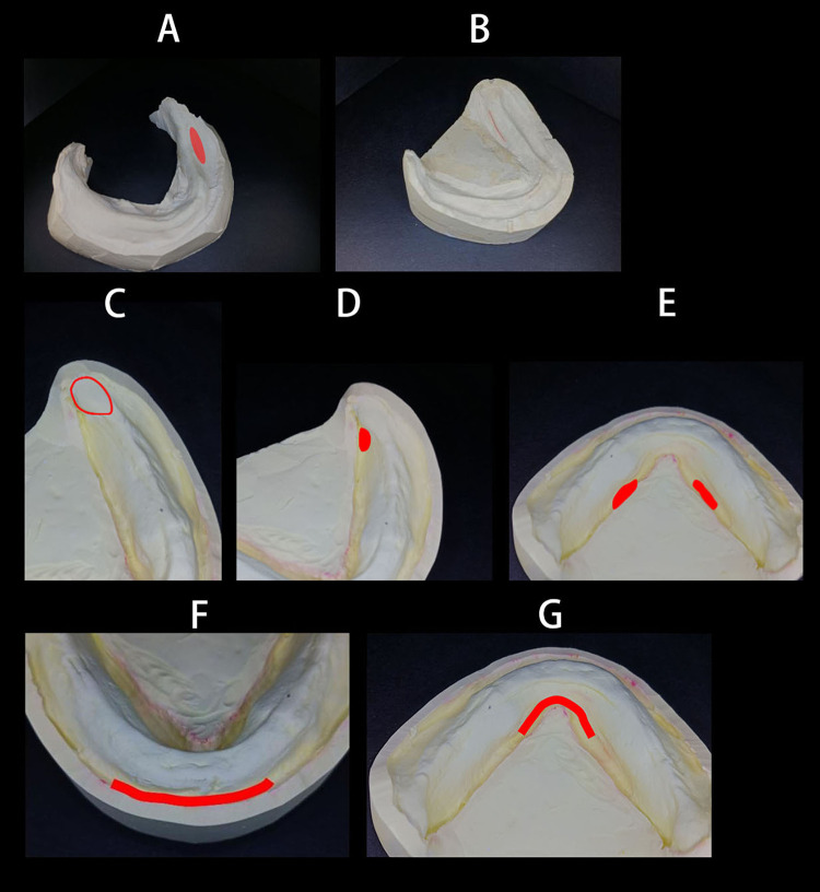
Results: Statistically significant differences were observed in three specific regions: the posterior fossa of the mylohyoid, the submandibular gland region, and the labial vestibule (p < 0.05). In contrast, differences in the posterior molar pad area were minimal (p > 0.05). Overall, the final model exhibited significant deviations from the initial model (p < 0.05), with the greatest variation observed in the posterior fossa of the mylohyoid and the least variation in the posterior molar pad area.
Conclusion: The final mandibular complete denture model, constructed using BPS, demonstrates improved accuracy in representing mucosal movement, shape, and positioning under occlusal pressure compared to the initial model. The posterior fossa of the mylohyoid, submandibular gland region, and labial vestibule exhibited enhanced delineation of mucosal movement boundaries, contributing to a more precise functional representation.
期刊介绍:
Therapeutics and Clinical Risk Management is an international, peer-reviewed journal of clinical therapeutics and risk management, focusing on concise rapid reporting of clinical studies in all therapeutic areas, outcomes, safety, and programs for the effective, safe, and sustained use of medicines, therapeutic and surgical interventions in all clinical areas.
The journal welcomes submissions covering original research, clinical and epidemiological studies, reviews, guidelines, expert opinion and commentary. The journal will consider case reports but only if they make a valuable and original contribution to the literature.
As of 18th March 2019, Therapeutics and Clinical Risk Management will no longer consider meta-analyses for publication.
The journal does not accept study protocols, animal-based or cell line-based studies.
 求助内容:
求助内容: 应助结果提醒方式:


