N. V. Antipova, D. A. Bondarenko, D. V. Mazur, A. A. Isakova, M. E. Gasparian, O. I. Patsap, V. M. Pavlov, E. S. Mikhailov, N. A. Goryacheva, D. I. Rzhevsky, S. G. Semushina, D. A. Dolgikh, A. N. Murashev, A. V. Yagolovich
{"title":"NSG小鼠颅内胶质母细胞瘤PDX模型的建立","authors":"N. V. Antipova, D. A. Bondarenko, D. V. Mazur, A. A. Isakova, M. E. Gasparian, O. I. Patsap, V. M. Pavlov, E. S. Mikhailov, N. A. Goryacheva, D. I. Rzhevsky, S. G. Semushina, D. A. Dolgikh, A. N. Murashev, A. V. Yagolovich","doi":"10.1134/S1068162025602514","DOIUrl":null,"url":null,"abstract":"<p><b>Objective:</b> Establishment of an orthotopic intracranial model from human glioblastoma cell culture in immunodeficient mice is important both for studying the invasiveness and aggressiveness of tumor cells and for developing a reliable model for assessing the effectiveness of new drugs for glioblastoma therapy. The aim of this work was to perform a comparative study of tumorigenicity of established and primary patient-derived glioblastoma cells during subcutaneous and orthotopic intracranial xenotransplantation into immunodeficient NSG mice. <b>Methods:</b> The glioblastoma cell line U87MG and primary patient-derived glioblastoma cells (022 culture) were characterized in terms of morphology and molecular subtype. The expression of specific markers was analyzed by RNA-seq. Cells were xenografted into immunodeficient NSG mice either subcutaneously or orthotopically into the corpus striatum. Tumors were examined using immunohistochemical staining for glial markers. <b>Results and Discussion:</b> It was shown that in both groups of animals with orthotopic xenografts, tumors grew both deep into the brain tissue and on the brain surface, and in the case of the primary 022 culture, growth toward the ventricles was also noted. Morphologically, primary 022 cells had an epithelioid morphology, while U87MG cells were more sarcomatoid. Importantly, the U87MG cell line was tumorigenic in both localizations. However, the primary 022 culture formed tumors only during intracranial, but not during subcutaneous xenotransplantation, which indicates the neurospecificity of this model. <b>Conclusions:</b> The primary patient-derived glioblastoma culture 022 may serve as a more relevant model of glioblastoma compared to the U87MG cell line-based model.</p>","PeriodicalId":758,"journal":{"name":"Russian Journal of Bioorganic Chemistry","volume":"51 5","pages":"2034 - 2040"},"PeriodicalIF":1.7000,"publicationDate":"2025-09-28","publicationTypes":"Journal Article","fieldsOfStudy":null,"isOpenAccess":false,"openAccessPdf":"","citationCount":"0","resultStr":"{\"title\":\"Development of an Experimental Intracranial PDX Model of Human Glioblastoma in NSG Mice\",\"authors\":\"N. V. Antipova, D. A. Bondarenko, D. V. Mazur, A. A. Isakova, M. E. Gasparian, O. I. Patsap, V. M. Pavlov, E. S. Mikhailov, N. A. Goryacheva, D. I. Rzhevsky, S. G. Semushina, D. A. Dolgikh, A. N. Murashev, A. V. Yagolovich\",\"doi\":\"10.1134/S1068162025602514\",\"DOIUrl\":null,\"url\":null,\"abstract\":\"<p><b>Objective:</b> Establishment of an orthotopic intracranial model from human glioblastoma cell culture in immunodeficient mice is important both for studying the invasiveness and aggressiveness of tumor cells and for developing a reliable model for assessing the effectiveness of new drugs for glioblastoma therapy. The aim of this work was to perform a comparative study of tumorigenicity of established and primary patient-derived glioblastoma cells during subcutaneous and orthotopic intracranial xenotransplantation into immunodeficient NSG mice. <b>Methods:</b> The glioblastoma cell line U87MG and primary patient-derived glioblastoma cells (022 culture) were characterized in terms of morphology and molecular subtype. The expression of specific markers was analyzed by RNA-seq. Cells were xenografted into immunodeficient NSG mice either subcutaneously or orthotopically into the corpus striatum. Tumors were examined using immunohistochemical staining for glial markers. <b>Results and Discussion:</b> It was shown that in both groups of animals with orthotopic xenografts, tumors grew both deep into the brain tissue and on the brain surface, and in the case of the primary 022 culture, growth toward the ventricles was also noted. Morphologically, primary 022 cells had an epithelioid morphology, while U87MG cells were more sarcomatoid. Importantly, the U87MG cell line was tumorigenic in both localizations. However, the primary 022 culture formed tumors only during intracranial, but not during subcutaneous xenotransplantation, which indicates the neurospecificity of this model. <b>Conclusions:</b> The primary patient-derived glioblastoma culture 022 may serve as a more relevant model of glioblastoma compared to the U87MG cell line-based model.</p>\",\"PeriodicalId\":758,\"journal\":{\"name\":\"Russian Journal of Bioorganic Chemistry\",\"volume\":\"51 5\",\"pages\":\"2034 - 2040\"},\"PeriodicalIF\":1.7000,\"publicationDate\":\"2025-09-28\",\"publicationTypes\":\"Journal Article\",\"fieldsOfStudy\":null,\"isOpenAccess\":false,\"openAccessPdf\":\"\",\"citationCount\":\"0\",\"resultStr\":null,\"platform\":\"Semanticscholar\",\"paperid\":null,\"PeriodicalName\":\"Russian Journal of Bioorganic Chemistry\",\"FirstCategoryId\":\"92\",\"ListUrlMain\":\"https://link.springer.com/article/10.1134/S1068162025602514\",\"RegionNum\":4,\"RegionCategory\":\"化学\",\"ArticlePicture\":[],\"TitleCN\":null,\"AbstractTextCN\":null,\"PMCID\":null,\"EPubDate\":\"\",\"PubModel\":\"\",\"JCR\":\"Q4\",\"JCRName\":\"BIOCHEMISTRY & MOLECULAR BIOLOGY\",\"Score\":null,\"Total\":0}","platform":"Semanticscholar","paperid":null,"PeriodicalName":"Russian Journal of Bioorganic Chemistry","FirstCategoryId":"92","ListUrlMain":"https://link.springer.com/article/10.1134/S1068162025602514","RegionNum":4,"RegionCategory":"化学","ArticlePicture":[],"TitleCN":null,"AbstractTextCN":null,"PMCID":null,"EPubDate":"","PubModel":"","JCR":"Q4","JCRName":"BIOCHEMISTRY & MOLECULAR BIOLOGY","Score":null,"Total":0}
Development of an Experimental Intracranial PDX Model of Human Glioblastoma in NSG Mice
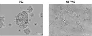
Objective: Establishment of an orthotopic intracranial model from human glioblastoma cell culture in immunodeficient mice is important both for studying the invasiveness and aggressiveness of tumor cells and for developing a reliable model for assessing the effectiveness of new drugs for glioblastoma therapy. The aim of this work was to perform a comparative study of tumorigenicity of established and primary patient-derived glioblastoma cells during subcutaneous and orthotopic intracranial xenotransplantation into immunodeficient NSG mice. Methods: The glioblastoma cell line U87MG and primary patient-derived glioblastoma cells (022 culture) were characterized in terms of morphology and molecular subtype. The expression of specific markers was analyzed by RNA-seq. Cells were xenografted into immunodeficient NSG mice either subcutaneously or orthotopically into the corpus striatum. Tumors were examined using immunohistochemical staining for glial markers. Results and Discussion: It was shown that in both groups of animals with orthotopic xenografts, tumors grew both deep into the brain tissue and on the brain surface, and in the case of the primary 022 culture, growth toward the ventricles was also noted. Morphologically, primary 022 cells had an epithelioid morphology, while U87MG cells were more sarcomatoid. Importantly, the U87MG cell line was tumorigenic in both localizations. However, the primary 022 culture formed tumors only during intracranial, but not during subcutaneous xenotransplantation, which indicates the neurospecificity of this model. Conclusions: The primary patient-derived glioblastoma culture 022 may serve as a more relevant model of glioblastoma compared to the U87MG cell line-based model.
期刊介绍:
Russian Journal of Bioorganic Chemistry publishes reviews and original experimental and theoretical studies on the structure, function, structure–activity relationships, and synthesis of biopolymers, such as proteins, nucleic acids, polysaccharides, mixed biopolymers, and their complexes, and low-molecular-weight biologically active compounds (peptides, sugars, lipids, antibiotics, etc.). The journal also covers selected aspects of neuro- and immunochemistry, biotechnology, and ecology.
 求助内容:
求助内容: 应助结果提醒方式:


