Melanie Scherer, Barbara Hochecker, Katja Matt, Alica Meßmer, Jörg Bergemann
{"title":"使用Guava®自噬和流式细胞术检测人外周血单核细胞的自噬。","authors":"Melanie Scherer, Barbara Hochecker, Katja Matt, Alica Meßmer, Jörg Bergemann","doi":"10.21769/BioProtoc.5457","DOIUrl":null,"url":null,"abstract":"<p><p>Autophagy plays a crucial role in cellular homeostasis and is responsible for removing and degrading damaged cytoplasmic cargo. This lysosome-mediated catabolic process removes defective organelles and misfolded proteins, and impaired autophagy has been directly linked to ageing and numerous diseases. This emphasises the importance of developing intervention methods to counteract this dysregulation. One promising intervention is thermal therapy, specifically hyperthermia, which is described in this protocol. In order to investigate this form of treatment, a rapid and reliable detection method is required to allow comparison of autophagy status under different conditions. While methods such as transmission electron microscopy (TEM) or western blotting can provide valuable structural analysis, they are often time-consuming and expensive, and are not suitable for small, round cells such as peripheral blood mononuclear cells (PBMCs). The method described in this protocol enables absolute quantification of PBMCs using the Guava<sup>®</sup> Autophagy Detection kit after heat treatment with water-filtered infrared-A radiation (wIRA), compared with an untreated control. This method is based on antibody labelling, and subsequent flow cytometric analysis enables the number of autophagosomes to be determined by measuring the FITC intensity. This protocol provides rapid, reliable results and can be adapted to investigate not only heat therapy, but also other interventions, such as caloric restriction. Key features • Rapid and reliable ex vivo quantification of autophagy in living cells. • Optimised protocol for the determination of autophagy in primary human blood cells. • Allows the testing of active substances and treatments concerning autophagy. • Flow cytometry-based method for the determination of autophagy.</p>","PeriodicalId":93907,"journal":{"name":"Bio-protocol","volume":"15 18","pages":"e5457"},"PeriodicalIF":1.1000,"publicationDate":"2025-09-20","publicationTypes":"Journal Article","fieldsOfStudy":null,"isOpenAccess":false,"openAccessPdf":"https://www.ncbi.nlm.nih.gov/pmc/articles/PMC12457839/pdf/","citationCount":"0","resultStr":"{\"title\":\"Detection of Autophagy in Human Peripheral Blood Mononuclear Cells Using Guava<sup>®</sup> Autophagy and Flow Cytometry.\",\"authors\":\"Melanie Scherer, Barbara Hochecker, Katja Matt, Alica Meßmer, Jörg Bergemann\",\"doi\":\"10.21769/BioProtoc.5457\",\"DOIUrl\":null,\"url\":null,\"abstract\":\"<p><p>Autophagy plays a crucial role in cellular homeostasis and is responsible for removing and degrading damaged cytoplasmic cargo. This lysosome-mediated catabolic process removes defective organelles and misfolded proteins, and impaired autophagy has been directly linked to ageing and numerous diseases. This emphasises the importance of developing intervention methods to counteract this dysregulation. One promising intervention is thermal therapy, specifically hyperthermia, which is described in this protocol. In order to investigate this form of treatment, a rapid and reliable detection method is required to allow comparison of autophagy status under different conditions. While methods such as transmission electron microscopy (TEM) or western blotting can provide valuable structural analysis, they are often time-consuming and expensive, and are not suitable for small, round cells such as peripheral blood mononuclear cells (PBMCs). The method described in this protocol enables absolute quantification of PBMCs using the Guava<sup>®</sup> Autophagy Detection kit after heat treatment with water-filtered infrared-A radiation (wIRA), compared with an untreated control. This method is based on antibody labelling, and subsequent flow cytometric analysis enables the number of autophagosomes to be determined by measuring the FITC intensity. This protocol provides rapid, reliable results and can be adapted to investigate not only heat therapy, but also other interventions, such as caloric restriction. Key features • Rapid and reliable ex vivo quantification of autophagy in living cells. • Optimised protocol for the determination of autophagy in primary human blood cells. • Allows the testing of active substances and treatments concerning autophagy. • Flow cytometry-based method for the determination of autophagy.</p>\",\"PeriodicalId\":93907,\"journal\":{\"name\":\"Bio-protocol\",\"volume\":\"15 18\",\"pages\":\"e5457\"},\"PeriodicalIF\":1.1000,\"publicationDate\":\"2025-09-20\",\"publicationTypes\":\"Journal Article\",\"fieldsOfStudy\":null,\"isOpenAccess\":false,\"openAccessPdf\":\"https://www.ncbi.nlm.nih.gov/pmc/articles/PMC12457839/pdf/\",\"citationCount\":\"0\",\"resultStr\":null,\"platform\":\"Semanticscholar\",\"paperid\":null,\"PeriodicalName\":\"Bio-protocol\",\"FirstCategoryId\":\"1085\",\"ListUrlMain\":\"https://doi.org/10.21769/BioProtoc.5457\",\"RegionNum\":0,\"RegionCategory\":null,\"ArticlePicture\":[],\"TitleCN\":null,\"AbstractTextCN\":null,\"PMCID\":null,\"EPubDate\":\"\",\"PubModel\":\"\",\"JCR\":\"Q3\",\"JCRName\":\"BIOLOGY\",\"Score\":null,\"Total\":0}","platform":"Semanticscholar","paperid":null,"PeriodicalName":"Bio-protocol","FirstCategoryId":"1085","ListUrlMain":"https://doi.org/10.21769/BioProtoc.5457","RegionNum":0,"RegionCategory":null,"ArticlePicture":[],"TitleCN":null,"AbstractTextCN":null,"PMCID":null,"EPubDate":"","PubModel":"","JCR":"Q3","JCRName":"BIOLOGY","Score":null,"Total":0}
Detection of Autophagy in Human Peripheral Blood Mononuclear Cells Using Guava® Autophagy and Flow Cytometry.
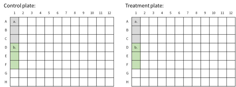
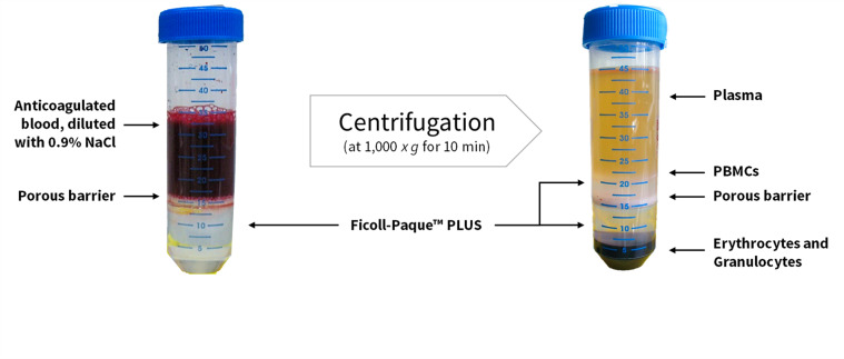
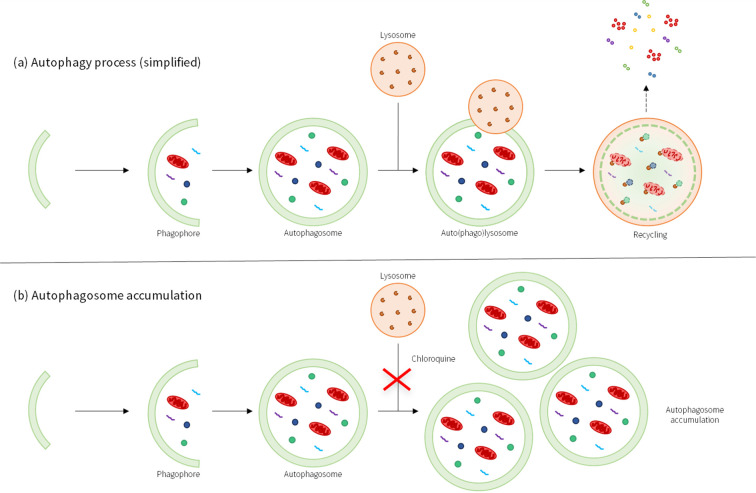
Autophagy plays a crucial role in cellular homeostasis and is responsible for removing and degrading damaged cytoplasmic cargo. This lysosome-mediated catabolic process removes defective organelles and misfolded proteins, and impaired autophagy has been directly linked to ageing and numerous diseases. This emphasises the importance of developing intervention methods to counteract this dysregulation. One promising intervention is thermal therapy, specifically hyperthermia, which is described in this protocol. In order to investigate this form of treatment, a rapid and reliable detection method is required to allow comparison of autophagy status under different conditions. While methods such as transmission electron microscopy (TEM) or western blotting can provide valuable structural analysis, they are often time-consuming and expensive, and are not suitable for small, round cells such as peripheral blood mononuclear cells (PBMCs). The method described in this protocol enables absolute quantification of PBMCs using the Guava® Autophagy Detection kit after heat treatment with water-filtered infrared-A radiation (wIRA), compared with an untreated control. This method is based on antibody labelling, and subsequent flow cytometric analysis enables the number of autophagosomes to be determined by measuring the FITC intensity. This protocol provides rapid, reliable results and can be adapted to investigate not only heat therapy, but also other interventions, such as caloric restriction. Key features • Rapid and reliable ex vivo quantification of autophagy in living cells. • Optimised protocol for the determination of autophagy in primary human blood cells. • Allows the testing of active substances and treatments concerning autophagy. • Flow cytometry-based method for the determination of autophagy.

 求助内容:
求助内容: 应助结果提醒方式:


