Kseniya V Nevskaya, Alexandra G Pershina, Lina V Efimova, Ekaterina V Sukhinina, Polina K Kozlova, Alina Yu Ryzhkova, Ekaterina S Hmelevskaya, Marina K Ibragimova, Irina A Tsydenova, Nikolai V Litviakov, Elena V Udut
{"title":"用于检测抗转移治疗的三维肝球体乳腺癌微转移模型。","authors":"Kseniya V Nevskaya, Alexandra G Pershina, Lina V Efimova, Ekaterina V Sukhinina, Polina K Kozlova, Alina Yu Ryzhkova, Ekaterina S Hmelevskaya, Marina K Ibragimova, Irina A Tsydenova, Nikolai V Litviakov, Elena V Udut","doi":"10.21769/BioProtoc.5454","DOIUrl":null,"url":null,"abstract":"<p><p>Even though the survival and proliferation stages of cancer cells that have newly settled at a metastatic site are the rate-limiting stages and the most promising targets for drugs, there is a lack of models of the earliest stage of metastasis formation. A method for modeling breast cancer liver metastasis is described here: a stage of transition of a differentiated tumor cell into a cell actively proliferating in a three-dimensional (3D) liver spheroid. Opposite to existing heterocellular 3D models of metastases, the protocol allows modeling the initial stage of liver colonization by metastatic cells, the so-called \"micrometastases.\" The method includes obtaining a line of fluorescent tumor cells, fluorescence-activated sorting of differentiated cells, preparing a single-cell suspension of liver cells, forming a liver spheroid in an agarose mold, inducing the tumor cell dedifferentiation and proliferation using IL-6, and intravital microscopy of spheroids, with subsequent processing and analysis of fluorescent images in the ImageJ software. The performance of the proposed model was demonstrated using microRNA therapeutics. The ability of a combination of microRNAs to suppress the transition of micrometastasis to macrometastasis in the 3D liver spheroid was confirmed by an immunofluorescent assay of spheroid sections and transcriptome analysis. Key features • The method introduces a 3D model of liver micrometastasis formation using differentiated tumor cells. • The 3D spheroid consists of all the main types of normal liver cells and better reproduces the microenvironment. • The method allows one to evaluate the effectiveness of a drug that blocks the transition of micrometastases to macrometastases. • The model is optimal for studying RNA-based therapeutic agents, as well as prodrugs that require metabolism in the liver for activation.</p>","PeriodicalId":93907,"journal":{"name":"Bio-protocol","volume":"15 18","pages":"e5454"},"PeriodicalIF":1.1000,"publicationDate":"2025-09-20","publicationTypes":"Journal Article","fieldsOfStudy":null,"isOpenAccess":false,"openAccessPdf":"https://www.ncbi.nlm.nih.gov/pmc/articles/PMC12457848/pdf/","citationCount":"0","resultStr":"{\"title\":\"A Model of Breast Cancer Micrometastasis in a Three-Dimensional (3D) Liver Spheroid for Testing an Antimetastatic Therapy.\",\"authors\":\"Kseniya V Nevskaya, Alexandra G Pershina, Lina V Efimova, Ekaterina V Sukhinina, Polina K Kozlova, Alina Yu Ryzhkova, Ekaterina S Hmelevskaya, Marina K Ibragimova, Irina A Tsydenova, Nikolai V Litviakov, Elena V Udut\",\"doi\":\"10.21769/BioProtoc.5454\",\"DOIUrl\":null,\"url\":null,\"abstract\":\"<p><p>Even though the survival and proliferation stages of cancer cells that have newly settled at a metastatic site are the rate-limiting stages and the most promising targets for drugs, there is a lack of models of the earliest stage of metastasis formation. A method for modeling breast cancer liver metastasis is described here: a stage of transition of a differentiated tumor cell into a cell actively proliferating in a three-dimensional (3D) liver spheroid. Opposite to existing heterocellular 3D models of metastases, the protocol allows modeling the initial stage of liver colonization by metastatic cells, the so-called \\\"micrometastases.\\\" The method includes obtaining a line of fluorescent tumor cells, fluorescence-activated sorting of differentiated cells, preparing a single-cell suspension of liver cells, forming a liver spheroid in an agarose mold, inducing the tumor cell dedifferentiation and proliferation using IL-6, and intravital microscopy of spheroids, with subsequent processing and analysis of fluorescent images in the ImageJ software. The performance of the proposed model was demonstrated using microRNA therapeutics. The ability of a combination of microRNAs to suppress the transition of micrometastasis to macrometastasis in the 3D liver spheroid was confirmed by an immunofluorescent assay of spheroid sections and transcriptome analysis. Key features • The method introduces a 3D model of liver micrometastasis formation using differentiated tumor cells. • The 3D spheroid consists of all the main types of normal liver cells and better reproduces the microenvironment. • The method allows one to evaluate the effectiveness of a drug that blocks the transition of micrometastases to macrometastases. • The model is optimal for studying RNA-based therapeutic agents, as well as prodrugs that require metabolism in the liver for activation.</p>\",\"PeriodicalId\":93907,\"journal\":{\"name\":\"Bio-protocol\",\"volume\":\"15 18\",\"pages\":\"e5454\"},\"PeriodicalIF\":1.1000,\"publicationDate\":\"2025-09-20\",\"publicationTypes\":\"Journal Article\",\"fieldsOfStudy\":null,\"isOpenAccess\":false,\"openAccessPdf\":\"https://www.ncbi.nlm.nih.gov/pmc/articles/PMC12457848/pdf/\",\"citationCount\":\"0\",\"resultStr\":null,\"platform\":\"Semanticscholar\",\"paperid\":null,\"PeriodicalName\":\"Bio-protocol\",\"FirstCategoryId\":\"1085\",\"ListUrlMain\":\"https://doi.org/10.21769/BioProtoc.5454\",\"RegionNum\":0,\"RegionCategory\":null,\"ArticlePicture\":[],\"TitleCN\":null,\"AbstractTextCN\":null,\"PMCID\":null,\"EPubDate\":\"\",\"PubModel\":\"\",\"JCR\":\"Q3\",\"JCRName\":\"BIOLOGY\",\"Score\":null,\"Total\":0}","platform":"Semanticscholar","paperid":null,"PeriodicalName":"Bio-protocol","FirstCategoryId":"1085","ListUrlMain":"https://doi.org/10.21769/BioProtoc.5454","RegionNum":0,"RegionCategory":null,"ArticlePicture":[],"TitleCN":null,"AbstractTextCN":null,"PMCID":null,"EPubDate":"","PubModel":"","JCR":"Q3","JCRName":"BIOLOGY","Score":null,"Total":0}
A Model of Breast Cancer Micrometastasis in a Three-Dimensional (3D) Liver Spheroid for Testing an Antimetastatic Therapy.
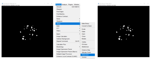
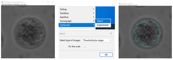
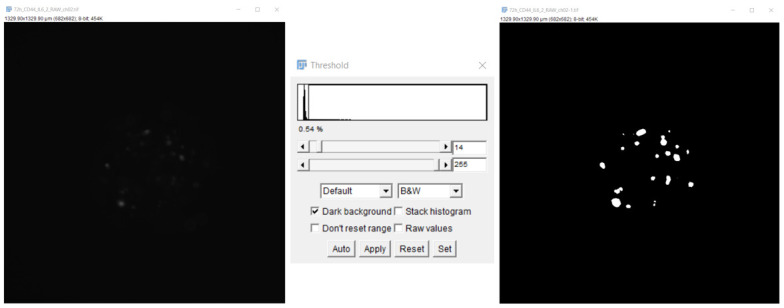
Even though the survival and proliferation stages of cancer cells that have newly settled at a metastatic site are the rate-limiting stages and the most promising targets for drugs, there is a lack of models of the earliest stage of metastasis formation. A method for modeling breast cancer liver metastasis is described here: a stage of transition of a differentiated tumor cell into a cell actively proliferating in a three-dimensional (3D) liver spheroid. Opposite to existing heterocellular 3D models of metastases, the protocol allows modeling the initial stage of liver colonization by metastatic cells, the so-called "micrometastases." The method includes obtaining a line of fluorescent tumor cells, fluorescence-activated sorting of differentiated cells, preparing a single-cell suspension of liver cells, forming a liver spheroid in an agarose mold, inducing the tumor cell dedifferentiation and proliferation using IL-6, and intravital microscopy of spheroids, with subsequent processing and analysis of fluorescent images in the ImageJ software. The performance of the proposed model was demonstrated using microRNA therapeutics. The ability of a combination of microRNAs to suppress the transition of micrometastasis to macrometastasis in the 3D liver spheroid was confirmed by an immunofluorescent assay of spheroid sections and transcriptome analysis. Key features • The method introduces a 3D model of liver micrometastasis formation using differentiated tumor cells. • The 3D spheroid consists of all the main types of normal liver cells and better reproduces the microenvironment. • The method allows one to evaluate the effectiveness of a drug that blocks the transition of micrometastases to macrometastases. • The model is optimal for studying RNA-based therapeutic agents, as well as prodrugs that require metabolism in the liver for activation.

 求助内容:
求助内容: 应助结果提醒方式:


