Neel Raja, Elias Petrou, Sonal Saran, Hasaam Uldin, Morgan Jones, Fahid Rasul, Kapil Shirodkar, Shashank Chapala, Rajesh Botchu
{"title":"评估后齿状突倾斜:考虑脊柱侧凸。","authors":"Neel Raja, Elias Petrou, Sonal Saran, Hasaam Uldin, Morgan Jones, Fahid Rasul, Kapil Shirodkar, Shashank Chapala, Rajesh Botchu","doi":"10.4103/jcvjs.jcvjs_89_25","DOIUrl":null,"url":null,"abstract":"<p><strong>Objective: </strong>The odontoid process is an important anatomical structure providing a balance of mobility and stability at the craniocervical junction, with structural and biomechanical associations, and morphology that can be quantified with various measurements. The odontoid tilt angle is a measurement that must be accurately performed and can guide further investigations.</p><p><strong>Materials and methods: </strong>Retrospective analysis of 100 cervical spinal magnetic resonance imaging was performed on patients investigated for neck pain, with a known history of scoliosis, and compared with 50 control patients. Posterior odontoid tilt and Cobb angles were measured by a musculoskeletal radiology fellow and a fellowship-trained musculoskeletal radiologist with more than 10 years of experience, with descriptive statistics then performed on the measurements.</p><p><strong>Results: </strong>One hundred and thirty-two patients met the inclusion criteria, across both the scoliosis and control groups. 9 (18%) patients from the control group demonstrated posterior odontoid tilt, compared with 35 (43%) of patients in the scoliosis group. A range of scoliosis curve morphologies were demonstrated: 62 thoracolumbar, 10 thoracic, 9 lumbar, and 1 cervicothoracic, with average Cobb angles of 24.3°, 26.9°, 23.4, and 54°, respectively. There was good interobserver agreement for both measurements and a statistically significant difference in the posterior odontoid tilt measurements between groups (99% confidence interval, <i>P</i> = 0.0064).</p><p><strong>Conclusion: </strong>We recommend opportunistically assessing for the posterior odontoid tilt (Leaning odontoid tower of BRUMES (Botchu; Raja Rasul; Uldin; Morgan;Elias; Sonal, Shashank, Shirodkar). In cases with a posterior tilt angle >5°, we recommend whole spine imaging to assess for scoliosis in the thoracolumbar spine.</p>","PeriodicalId":51721,"journal":{"name":"Journal of Craniovertebral Junction and Spine","volume":"16 3","pages":"278-283"},"PeriodicalIF":1.3000,"publicationDate":"2025-07-01","publicationTypes":"Journal Article","fieldsOfStudy":null,"isOpenAccess":false,"openAccessPdf":"https://www.ncbi.nlm.nih.gov/pmc/articles/PMC12459940/pdf/","citationCount":"0","resultStr":"{\"title\":\"Assessment of posterior odontoid tilt: Think scoliosis.\",\"authors\":\"Neel Raja, Elias Petrou, Sonal Saran, Hasaam Uldin, Morgan Jones, Fahid Rasul, Kapil Shirodkar, Shashank Chapala, Rajesh Botchu\",\"doi\":\"10.4103/jcvjs.jcvjs_89_25\",\"DOIUrl\":null,\"url\":null,\"abstract\":\"<p><strong>Objective: </strong>The odontoid process is an important anatomical structure providing a balance of mobility and stability at the craniocervical junction, with structural and biomechanical associations, and morphology that can be quantified with various measurements. The odontoid tilt angle is a measurement that must be accurately performed and can guide further investigations.</p><p><strong>Materials and methods: </strong>Retrospective analysis of 100 cervical spinal magnetic resonance imaging was performed on patients investigated for neck pain, with a known history of scoliosis, and compared with 50 control patients. Posterior odontoid tilt and Cobb angles were measured by a musculoskeletal radiology fellow and a fellowship-trained musculoskeletal radiologist with more than 10 years of experience, with descriptive statistics then performed on the measurements.</p><p><strong>Results: </strong>One hundred and thirty-two patients met the inclusion criteria, across both the scoliosis and control groups. 9 (18%) patients from the control group demonstrated posterior odontoid tilt, compared with 35 (43%) of patients in the scoliosis group. A range of scoliosis curve morphologies were demonstrated: 62 thoracolumbar, 10 thoracic, 9 lumbar, and 1 cervicothoracic, with average Cobb angles of 24.3°, 26.9°, 23.4, and 54°, respectively. There was good interobserver agreement for both measurements and a statistically significant difference in the posterior odontoid tilt measurements between groups (99% confidence interval, <i>P</i> = 0.0064).</p><p><strong>Conclusion: </strong>We recommend opportunistically assessing for the posterior odontoid tilt (Leaning odontoid tower of BRUMES (Botchu; Raja Rasul; Uldin; Morgan;Elias; Sonal, Shashank, Shirodkar). In cases with a posterior tilt angle >5°, we recommend whole spine imaging to assess for scoliosis in the thoracolumbar spine.</p>\",\"PeriodicalId\":51721,\"journal\":{\"name\":\"Journal of Craniovertebral Junction and Spine\",\"volume\":\"16 3\",\"pages\":\"278-283\"},\"PeriodicalIF\":1.3000,\"publicationDate\":\"2025-07-01\",\"publicationTypes\":\"Journal Article\",\"fieldsOfStudy\":null,\"isOpenAccess\":false,\"openAccessPdf\":\"https://www.ncbi.nlm.nih.gov/pmc/articles/PMC12459940/pdf/\",\"citationCount\":\"0\",\"resultStr\":null,\"platform\":\"Semanticscholar\",\"paperid\":null,\"PeriodicalName\":\"Journal of Craniovertebral Junction and Spine\",\"FirstCategoryId\":\"1085\",\"ListUrlMain\":\"https://doi.org/10.4103/jcvjs.jcvjs_89_25\",\"RegionNum\":0,\"RegionCategory\":null,\"ArticlePicture\":[],\"TitleCN\":null,\"AbstractTextCN\":null,\"PMCID\":null,\"EPubDate\":\"2025/9/18 0:00:00\",\"PubModel\":\"Epub\",\"JCR\":\"Q2\",\"JCRName\":\"OTORHINOLARYNGOLOGY\",\"Score\":null,\"Total\":0}","platform":"Semanticscholar","paperid":null,"PeriodicalName":"Journal of Craniovertebral Junction and Spine","FirstCategoryId":"1085","ListUrlMain":"https://doi.org/10.4103/jcvjs.jcvjs_89_25","RegionNum":0,"RegionCategory":null,"ArticlePicture":[],"TitleCN":null,"AbstractTextCN":null,"PMCID":null,"EPubDate":"2025/9/18 0:00:00","PubModel":"Epub","JCR":"Q2","JCRName":"OTORHINOLARYNGOLOGY","Score":null,"Total":0}
Assessment of posterior odontoid tilt: Think scoliosis.
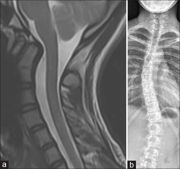
Objective: The odontoid process is an important anatomical structure providing a balance of mobility and stability at the craniocervical junction, with structural and biomechanical associations, and morphology that can be quantified with various measurements. The odontoid tilt angle is a measurement that must be accurately performed and can guide further investigations.
Materials and methods: Retrospective analysis of 100 cervical spinal magnetic resonance imaging was performed on patients investigated for neck pain, with a known history of scoliosis, and compared with 50 control patients. Posterior odontoid tilt and Cobb angles were measured by a musculoskeletal radiology fellow and a fellowship-trained musculoskeletal radiologist with more than 10 years of experience, with descriptive statistics then performed on the measurements.
Results: One hundred and thirty-two patients met the inclusion criteria, across both the scoliosis and control groups. 9 (18%) patients from the control group demonstrated posterior odontoid tilt, compared with 35 (43%) of patients in the scoliosis group. A range of scoliosis curve morphologies were demonstrated: 62 thoracolumbar, 10 thoracic, 9 lumbar, and 1 cervicothoracic, with average Cobb angles of 24.3°, 26.9°, 23.4, and 54°, respectively. There was good interobserver agreement for both measurements and a statistically significant difference in the posterior odontoid tilt measurements between groups (99% confidence interval, P = 0.0064).
Conclusion: We recommend opportunistically assessing for the posterior odontoid tilt (Leaning odontoid tower of BRUMES (Botchu; Raja Rasul; Uldin; Morgan;Elias; Sonal, Shashank, Shirodkar). In cases with a posterior tilt angle >5°, we recommend whole spine imaging to assess for scoliosis in the thoracolumbar spine.


 求助内容:
求助内容: 应助结果提醒方式:


