Christopher Schuppert, Stefanie Rahn, Nikolas D Schnellbächer, Frank Bergner, Michael Grass, Hans-Ulrich Kauczor, Stephan Skornitzke, Tim F Weber, Thuy D Do
{"title":"一种深度学习重建方法在双层检测CT系统临床胸腹骨盆扫描中的表现。","authors":"Christopher Schuppert, Stefanie Rahn, Nikolas D Schnellbächer, Frank Bergner, Michael Grass, Hans-Ulrich Kauczor, Stephan Skornitzke, Tim F Weber, Thuy D Do","doi":"10.3390/tomography11090094","DOIUrl":null,"url":null,"abstract":"<p><p><i>Objective</i>: The objective of this study was to compare the performance and robustness of a deep learning reconstruction method against established alternatives for soft tissue CT image reconstruction. <i>Materials and Methods</i>: Images were generated from portal venous phase chest-abdomen-pelvis CT scans (<i>n</i> = 99) acquired on a dual-layer spectral detector CT using filtered back projection, iterative model reconstruction (IMR), and deep learning reconstruction (DLR) with three parameter settings, namely 'standard', 'sharper', and 'smoother'. Experienced raters performed a quantitative assessment by considering attenuation stability and image noise levels in ten representative structures across all reconstruction methods, as well as a qualitative assessment using a four-point Likert scale (1 = poor, 2 = fair, 3 = good, 4 = excellent) for their overall perception of 'smoother' DLR and IMR images. One scan was excluded due to cachexia, which limited the quantitative measurements. <i>Results</i>: The inter-rater reliability for quantitative measurements ranged from moderate to excellent (<i>r</i> = 0.63-0.96). Attenuation values did not differ significantly between reconstruction methods except for DLR against IMR in the psoas muscle (mean + 3.0 HU, <i>p</i> < 0.001). Image noise levels differed significantly between reconstruction methods for all structures (all <i>p</i> < 0.001) and were lower than FBP with any DLR parameter setting. Image noise levels with 'smoother' DLR were predominantly lower than or equal to IMR, while they were higher with 'standard' DLR and 'sharper' DLR. The 'smoother' DLR images received a higher mean rating for overall image quality than the IMR images (3.7 vs. 2.3, <i>p</i> < 0.001). <i>Conclusions</i>: 'Smoother' DLR images were perceived by experienced readers as having improved quality compared to FBP and IMR while also exhibiting objectively lower or equivalent noise levels.</p>","PeriodicalId":51330,"journal":{"name":"Tomography","volume":"11 9","pages":""},"PeriodicalIF":2.2000,"publicationDate":"2025-08-25","publicationTypes":"Journal Article","fieldsOfStudy":null,"isOpenAccess":false,"openAccessPdf":"https://www.ncbi.nlm.nih.gov/pmc/articles/PMC12473457/pdf/","citationCount":"0","resultStr":"{\"title\":\"Performance of a Deep Learning Reconstruction Method on Clinical Chest-Abdomen-Pelvis Scans from a Dual-Layer Detector CT System.\",\"authors\":\"Christopher Schuppert, Stefanie Rahn, Nikolas D Schnellbächer, Frank Bergner, Michael Grass, Hans-Ulrich Kauczor, Stephan Skornitzke, Tim F Weber, Thuy D Do\",\"doi\":\"10.3390/tomography11090094\",\"DOIUrl\":null,\"url\":null,\"abstract\":\"<p><p><i>Objective</i>: The objective of this study was to compare the performance and robustness of a deep learning reconstruction method against established alternatives for soft tissue CT image reconstruction. <i>Materials and Methods</i>: Images were generated from portal venous phase chest-abdomen-pelvis CT scans (<i>n</i> = 99) acquired on a dual-layer spectral detector CT using filtered back projection, iterative model reconstruction (IMR), and deep learning reconstruction (DLR) with three parameter settings, namely 'standard', 'sharper', and 'smoother'. Experienced raters performed a quantitative assessment by considering attenuation stability and image noise levels in ten representative structures across all reconstruction methods, as well as a qualitative assessment using a four-point Likert scale (1 = poor, 2 = fair, 3 = good, 4 = excellent) for their overall perception of 'smoother' DLR and IMR images. One scan was excluded due to cachexia, which limited the quantitative measurements. <i>Results</i>: The inter-rater reliability for quantitative measurements ranged from moderate to excellent (<i>r</i> = 0.63-0.96). Attenuation values did not differ significantly between reconstruction methods except for DLR against IMR in the psoas muscle (mean + 3.0 HU, <i>p</i> < 0.001). Image noise levels differed significantly between reconstruction methods for all structures (all <i>p</i> < 0.001) and were lower than FBP with any DLR parameter setting. Image noise levels with 'smoother' DLR were predominantly lower than or equal to IMR, while they were higher with 'standard' DLR and 'sharper' DLR. The 'smoother' DLR images received a higher mean rating for overall image quality than the IMR images (3.7 vs. 2.3, <i>p</i> < 0.001). <i>Conclusions</i>: 'Smoother' DLR images were perceived by experienced readers as having improved quality compared to FBP and IMR while also exhibiting objectively lower or equivalent noise levels.</p>\",\"PeriodicalId\":51330,\"journal\":{\"name\":\"Tomography\",\"volume\":\"11 9\",\"pages\":\"\"},\"PeriodicalIF\":2.2000,\"publicationDate\":\"2025-08-25\",\"publicationTypes\":\"Journal Article\",\"fieldsOfStudy\":null,\"isOpenAccess\":false,\"openAccessPdf\":\"https://www.ncbi.nlm.nih.gov/pmc/articles/PMC12473457/pdf/\",\"citationCount\":\"0\",\"resultStr\":null,\"platform\":\"Semanticscholar\",\"paperid\":null,\"PeriodicalName\":\"Tomography\",\"FirstCategoryId\":\"3\",\"ListUrlMain\":\"https://doi.org/10.3390/tomography11090094\",\"RegionNum\":4,\"RegionCategory\":\"医学\",\"ArticlePicture\":[],\"TitleCN\":null,\"AbstractTextCN\":null,\"PMCID\":null,\"EPubDate\":\"\",\"PubModel\":\"\",\"JCR\":\"Q2\",\"JCRName\":\"RADIOLOGY, NUCLEAR MEDICINE & MEDICAL IMAGING\",\"Score\":null,\"Total\":0}","platform":"Semanticscholar","paperid":null,"PeriodicalName":"Tomography","FirstCategoryId":"3","ListUrlMain":"https://doi.org/10.3390/tomography11090094","RegionNum":4,"RegionCategory":"医学","ArticlePicture":[],"TitleCN":null,"AbstractTextCN":null,"PMCID":null,"EPubDate":"","PubModel":"","JCR":"Q2","JCRName":"RADIOLOGY, NUCLEAR MEDICINE & MEDICAL IMAGING","Score":null,"Total":0}
引用次数: 0
摘要
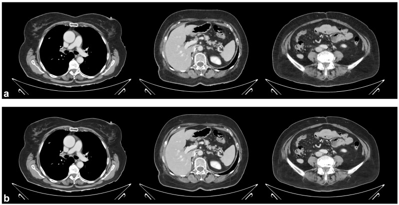
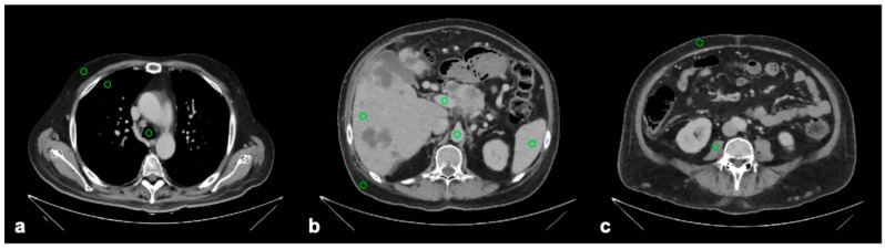
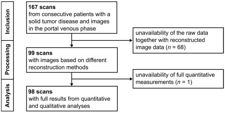
目的:本研究的目的是比较一种深度学习重建方法与现有的软组织CT图像重建方法的性能和鲁棒性。材料和方法:采用滤波后反投影、迭代模型重建(IMR)和深度学习重建(DLR),通过“标准”、“更清晰”和“更平滑”三个参数设置,对双层光谱检测器CT采集的门静脉期胸腹骨盆CT扫描(n = 99)进行图像生成。经验丰富的评分者通过考虑所有重建方法中十个代表性结构的衰减稳定性和图像噪声水平进行定量评估,并使用四点李克特量表(1 =差,2 =一般,3 =好,4 =优秀)对他们对“更平滑”的DLR和IMR图像的整体感知进行定性评估。一次扫描由于恶病质而被排除,这限制了定量测量。结果:定量测量的量表间信度为中等至优异(r = 0.63-0.96)。除腰大肌DLR与IMR的衰减值(平均+ 3.0 HU, p < 0.001)外,不同重建方法之间的衰减值无显著差异。图像噪声水平在所有结构的重建方法之间存在显著差异(均p < 0.001),并且在任何DLR参数设置下均低于FBP。“平滑”DLR的图像噪点水平主要低于或等于IMR,而“标准”DLR和“锐利”DLR的图像噪点水平则更高。“更平滑”的DLR图像在整体图像质量方面的平均评分高于IMR图像(3.7 vs. 2.3, p < 0.001)。结论:与FBP和IMR相比,经验丰富的读者认为“更平滑”的DLR图像质量更好,同时客观上也表现出更低或相当的噪声水平。
Performance of a Deep Learning Reconstruction Method on Clinical Chest-Abdomen-Pelvis Scans from a Dual-Layer Detector CT System.
Objective: The objective of this study was to compare the performance and robustness of a deep learning reconstruction method against established alternatives for soft tissue CT image reconstruction. Materials and Methods: Images were generated from portal venous phase chest-abdomen-pelvis CT scans (n = 99) acquired on a dual-layer spectral detector CT using filtered back projection, iterative model reconstruction (IMR), and deep learning reconstruction (DLR) with three parameter settings, namely 'standard', 'sharper', and 'smoother'. Experienced raters performed a quantitative assessment by considering attenuation stability and image noise levels in ten representative structures across all reconstruction methods, as well as a qualitative assessment using a four-point Likert scale (1 = poor, 2 = fair, 3 = good, 4 = excellent) for their overall perception of 'smoother' DLR and IMR images. One scan was excluded due to cachexia, which limited the quantitative measurements. Results: The inter-rater reliability for quantitative measurements ranged from moderate to excellent (r = 0.63-0.96). Attenuation values did not differ significantly between reconstruction methods except for DLR against IMR in the psoas muscle (mean + 3.0 HU, p < 0.001). Image noise levels differed significantly between reconstruction methods for all structures (all p < 0.001) and were lower than FBP with any DLR parameter setting. Image noise levels with 'smoother' DLR were predominantly lower than or equal to IMR, while they were higher with 'standard' DLR and 'sharper' DLR. The 'smoother' DLR images received a higher mean rating for overall image quality than the IMR images (3.7 vs. 2.3, p < 0.001). Conclusions: 'Smoother' DLR images were perceived by experienced readers as having improved quality compared to FBP and IMR while also exhibiting objectively lower or equivalent noise levels.
TomographyMedicine-Radiology, Nuclear Medicine and Imaging
CiteScore
2.70
自引率
10.50%
发文量
222
期刊介绍:
TomographyTM publishes basic (technical and pre-clinical) and clinical scientific articles which involve the advancement of imaging technologies. Tomography encompasses studies that use single or multiple imaging modalities including for example CT, US, PET, SPECT, MR and hyperpolarization technologies, as well as optical modalities (i.e. bioluminescence, photoacoustic, endomicroscopy, fiber optic imaging and optical computed tomography) in basic sciences, engineering, preclinical and clinical medicine.
Tomography also welcomes studies involving exploration and refinement of contrast mechanisms and image-derived metrics within and across modalities toward the development of novel imaging probes for image-based feedback and intervention. The use of imaging in biology and medicine provides unparalleled opportunities to noninvasively interrogate tissues to obtain real-time dynamic and quantitative information required for diagnosis and response to interventions and to follow evolving pathological conditions. As multi-modal studies and the complexities of imaging technologies themselves are ever increasing to provide advanced information to scientists and clinicians.
Tomography provides a unique publication venue allowing investigators the opportunity to more precisely communicate integrated findings related to the diverse and heterogeneous features associated with underlying anatomical, physiological, functional, metabolic and molecular genetic activities of normal and diseased tissue. Thus Tomography publishes peer-reviewed articles which involve the broad use of imaging of any tissue and disease type including both preclinical and clinical investigations. In addition, hardware/software along with chemical and molecular probe advances are welcome as they are deemed to significantly contribute towards the long-term goal of improving the overall impact of imaging on scientific and clinical discovery.

 求助内容:
求助内容: 应助结果提醒方式:


